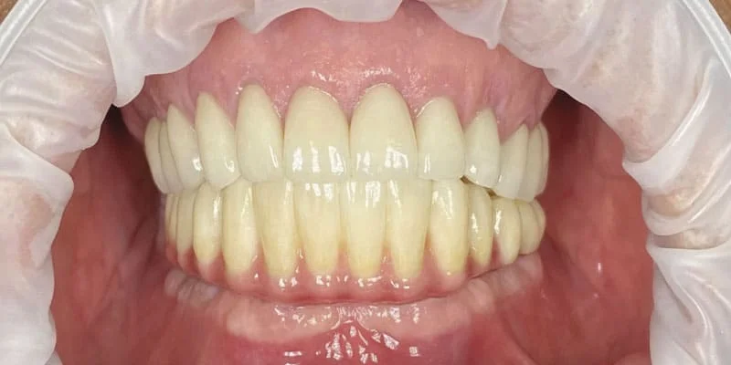
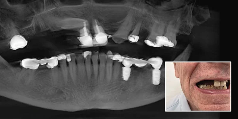
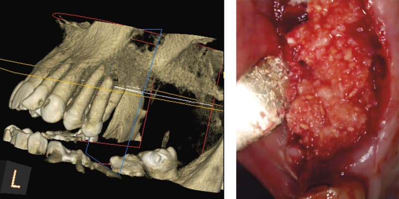
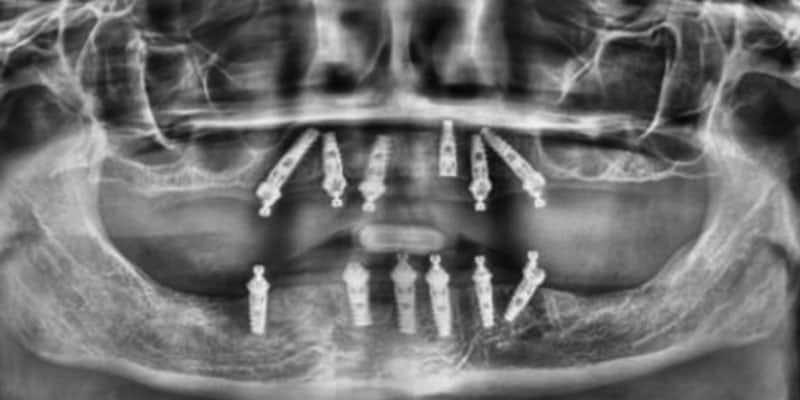
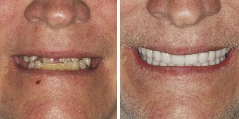
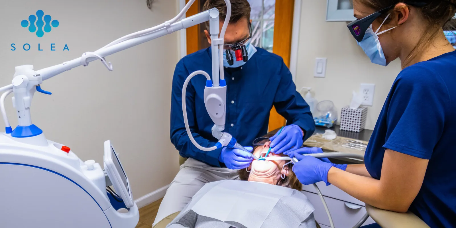
Dr. Robert Hayes discusses a predictable novel treatment modality for treating failing dentitions, edentulous, and potentially edentulous patients
Based on studies from many authors over 5 decades (Lopes, et al., 2106; Drago, 2016; Meloni, et al., 2017; Chan and Holmes, 2015; Patzelt, et al., 2014; Francetti, et al., 2008), this article is written from long clinical experience and deep understanding of the biomechanics of immediate load full arch treatment to emphasize and establish important parameters for successful technique (Holtzclaw, 2016).
Educational aims and objectives
This article aims to explain the benefits of and protocols for successful immediate load full arch treatment planning.
Expected outcomes
Implant Practice US subscribers can answer the CE questions to earn 2 hours of CE from reading this article. Correctly answering the questions will demonstrate the reader can:
- Read about this protocol and be aware of the evidence base supporting it.
- Read about some history of the full arch implant.
- Identify some aspects of prosthesis design.
- Recognize some aspects of angulation and load that facilitate successful outcomes.
- Identify the process for establishing the shelf.
- Realize how to determine access position.
- Realize how to guard against destructive bruxism.
Following careful investigation of the patient’s medical history and a tooth-by-tooth analysis, a detailed discussion that covers all treatment options — from living with gaps to wearing removable dentures or utilizing natural teeth as fixed bridge abutments or supplementing with sectional implant prosthetics — must also cover the anticipated scenarios and lifelong costs associated with each option.
Analyzing these cases is rather like high-level chess — how many strong pieces should be sacrificed early in order to win the long game?
Full arch implant treatment
Full arch implant treatment has a number of attractions. By eliminating the teeth, it removes the risks of decay, pulp necrosis, and periodontal infection. The first case was undertaken in 1965 by Dr. Per-Ingvar Brånemark, and many studies report the unassailable long-term success rates. The implants are linked by a supragingival metal bar providing cross-arch bracing and splinting, so no implant is ever bearing load alone. Full arch treatment is also an opportunity to establish an idealized version of the patient’s occlusion with canine rise/group function, balance, and harmony while eliminating destructive features such as cross-arch interference.
Since the 1960s, it has been recognized how a high-quality acrylic occlusion can protect the metal elements and bone from overloading as it softens impact and wears harmoniously, preventing the development of occlusal interferences (Brånemark, et al., 1977).
Research has also shown the development of hemidesmosomal attachment to acrylic, which we now utilize to control bacterial ingress and protect the bone from peri-implantitis (Listgarten and Lai, 1975). Ongoing investigations are evaluating the effects of incorporating antimicrobial and antibiotic agents into the prosthesis (Nazirkar, et al., 2014).
The long established adsorption of chlorhexidine gluconate onto acrylic supports transferring the prosthesis from the laboratory in such a solution and establishing a home-care regime that will refresh the anti-microbial reservoir (Bonesvoll and Olsen, 1974).
Prosthesis design
Since postoperative resorption of bone is inevitable, this author favors replacing the initial prosthesis at 4 months with a milled-titanium acrylic wrap that can be relined for years, with the original acrylic bridge being refurbished on the definitive models to provide a spare during laboratory procedures. The model ridge is trimmed by 1 millimeter to provide controlled gingival compression and development of a gingival trough in intimate contact with the dense, highly polished convex underside of the prosthesis. High-quality pressure-formed acrylic is used with great care on choice of fully chemically compatible teeth, metal prime, bond, mechanical retention on the bar and the teeth to achieve strength and durability. The esthetic result from high-quality acrylic work is far superior and easier to achieve than any other ceramic or composite alternative, and reparations can be much more readily accomplished.
Angulation and load
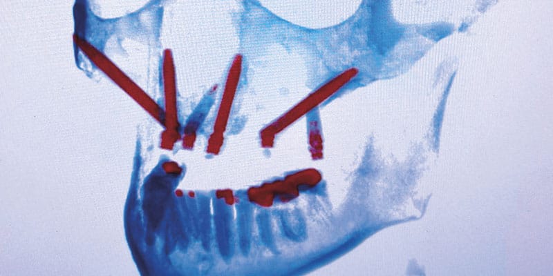
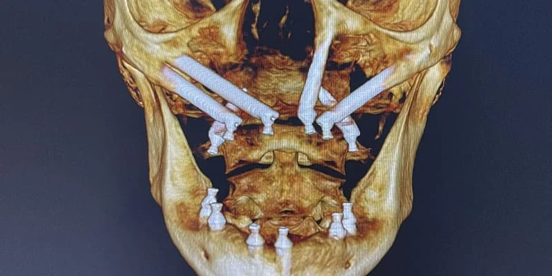
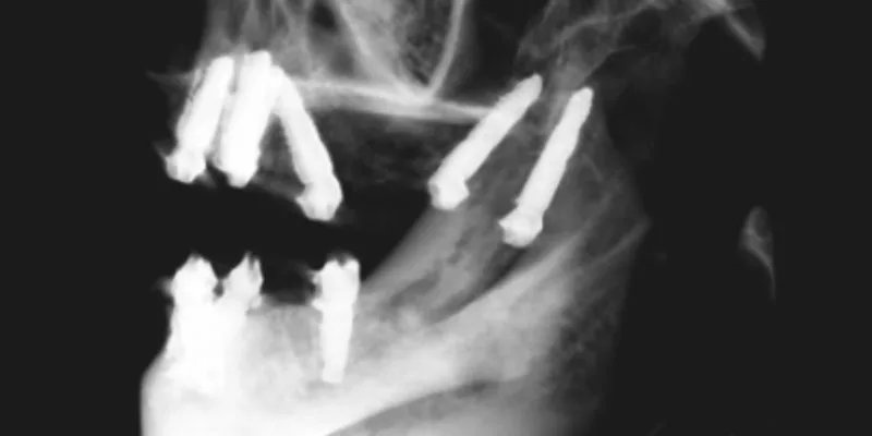
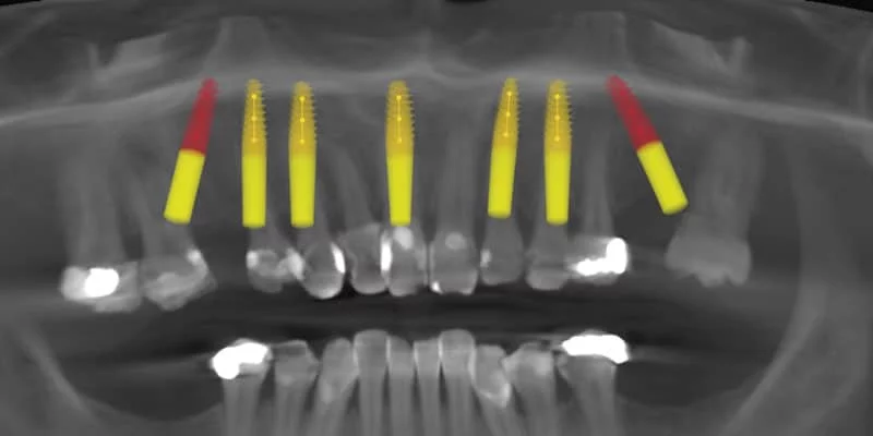
Biomechanical research has clearly demonstrated the advantage of placing long tilted implants to resist the high posterior load in the human jaw. Since the human jaw is a Class 3 lever (as is a nutcracker), the highest forces are always on the posterior implants (Lopes, et al., 2016; Silva, et al., 2010; Agliardi, et al., 2010). Thus placing short upright posterior implants below the sinus or above the nerve is inviting failure. To understand something of this, imagine a high occlusal point force on a single vertical implant — the bone contact surface has to bear all the force directly to prevent the implant being driven in like a nail.
With a 45º tilted implant, imagine the bone providing only a midpoint support. The force on the abutment would push the abutment end down, and the apical end would rise. Actually, the implant doesn’t move because the bone below the abutment end provides resistance, just as the bone above the apical end prevents the tilt. The forces are equal and opposite, so the load at such a point on the bone is halved.
None of the force applied during function — or more importantly parafunction — is directly along the long axis of the implant, so the resultant vectors of force along this natural path of movement of the implant are minimal. This angulation contributes to the near 99% implant success rates repeatedly reported, and it is incumbent on surgeons to apply this knowledge to full arch cases (Bhering, et al., 2016; Afrashtehfar, 2016; Saleh Saber, et al., 2015; Li, et al., 2015; Khatami and Smith, 2008).
The anatomy of the maxillary sinus and the mental nerve also lends itself to providing a resistant cortical surface immediately distal to the distal tilted implant, further enhancing its use, along with the widely understood benefit of reducing the cantilever and increasing the anterior-posterior spread of the abutments.
In extreme cases, it will be necessary to cross the anterior aspect of the sinus, place a zygomatic implant, or provide a posterior tilt in the opposite direction into the pterygoid plate. The most extreme maxillary cases are treated with quad zygoma, but in the mandible almost all can be treated with intra foraminal All-on-4 (All-on-3 in extreme cases) (De Moraes, et al., 2016; Jensen, et al., 2014; Malo, et al., 2013; Bedrossian, 2011; Ferreira, et al., 2010).
Application in the maxilla
The All-on-4 concept is absolutely applicable to the maxilla, but it must be recognized that the original papers were specific to a population of edentulous Portuguese pensioners accepting of a short arch concept and a 10-year survival (Malo, et al., 2011). When applying the concept to immediate placement in younger individuals with greater demands, it is wise to take into account the additional factors, including the lower density and reduced reliability of maxillary bone.
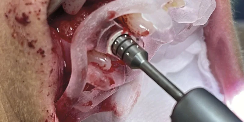
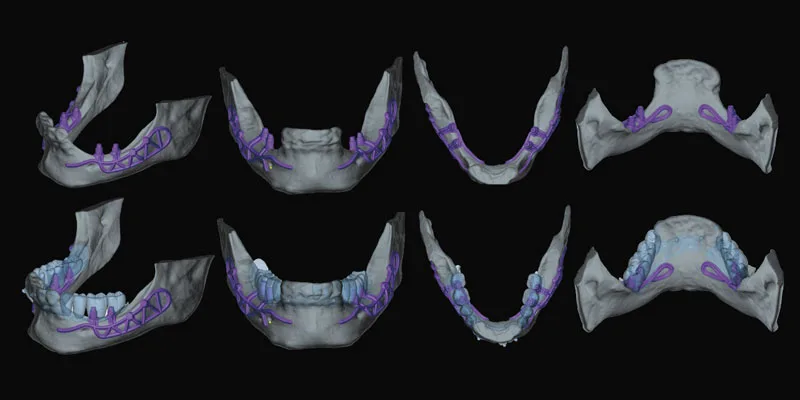
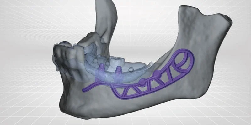
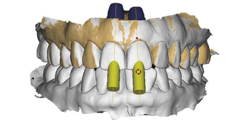
In many cases, the classic two tilted posterior and two anterior vertical implants should be supplemented by a third or fourth anterior implant and immediate load applied to all cases so that the implants are immediately splinted (Tallarico, et al., 2016; Silva, et al., 2010; Pomares, 2009). Placement of the biomechanically derived implant array must be prosthetically driven using a clear acrylic duplicate stent that informs positioning in all three dimensions.
This approach is derived from treatment of longstanding edentulous cases where fully guided surgery is a viable option using clear stereolithographically produced CAD/CAM devices produced digitally from CBCT planning software. In immediate cases, the dimensions of the surgical field are altered by reflection of full arch flaps, removal of teeth, roots and a sufficient depth of bone to accommodate the prosthesis (Faeghi Nejad, 2016; Butura, 2011; Pomares, 2009).
Establishing the shelf
It is vital to assess all aspects of the smile at the records appointment to determine the correct level of the transition line between the artificial and real gingivae, so that this is never visible in any expression of the lips, either anteriorly or within the buccal corridor.
The consequent requirement for ridge reduction must be communicated to the technician to inform the initial model ridge trimming, which must be flat rather than socketed to create a smooth, cleansable prosthetic junction (Abi Nader, et al., 2015; Krenmair, et al., 2014). The average case will require 3 millimeters of bone removal in the region of the extraction sockets — more if a gummy smile is to be corrected, and less if there has been significant bone loss due to severe periodontitis or a longstanding area of edentulous ridge (Jensen, et al., 2011a; Jensen, et al., 2010).
Access position
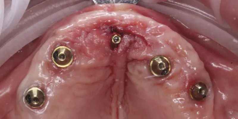
As each implant approaches final torque, account needs to be taken of the rotational position of the internal flats as these will determine the eventual access position of the prosthetic screws, which ideally occurs in the cingulum of the anterior teeth and the occlusal surface of the premolar or molar. Any facial deviation from this will risk an unsightly buccal access, and any lingual or palatal deviation will adversely affect the bulk of the prosthesis.
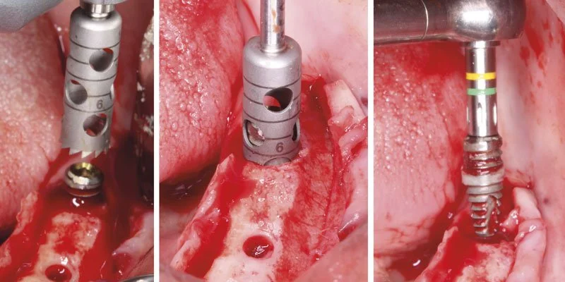
Some surgeons find it easier to use an external hex implant to directly observe the flats, but most manufacturers provide implant placement devices marked to indicate the internal flats. With the common hex design, this gives six positions for placing a 17º or 30º degree multi-unit abutment, three of which will be potentially restorable, giving a facial, mid, and lingual choice to abutment insertion, which should be chosen with reference to the clear stent by carefully referencing it to the final restorative position immediately prior to abutment connection.
It is wise to initially slightly under-prepare the osteotomies so that high primary stability (35-65 Ncm) of each implant is produced as the final rotational position is achieved, and abutments should be tightened to 15 Ncm, confirming full seating, which may require bone removal around any subcrestal aspect of the implant (Babbush, 2011; Jensen, et al., 2011b; Butura, 2011).
Guarding against bruxism
It should be recognized that many patients presenting for immediate full arch have destructive bruxism (De Rossi, et al., 2014). This is often apparent as a battle between the upper and lower teeth with units failing until eventually there are no opposing teeth in contact.
The pattern varies but, for example, the upper right and lower left buccal segments may be edentulous; then the lower incisors fail, and eventually the upper canines. Each loss depletes the dentition, and posterior loss in particular limits the parafunctional force. The “bruxism center” of the brain is searching for satisfying feedback and immediately introduces high forces to the transitional prosthesis. Creation of a protective, minimal overbite, minimal overjet, and idealized, close to edge-to-edge occlusion limited to first molars will protect the screws and implants from overloading.
Tightening abutments to 15 Ncm gives a positive fit, which will hold in most circumstances but release with more active bruxism — immediately protecting the implant bone attachment and alerting the clinician at the 2-month review. Protective steps can then be taken: retightening the screw to 10 Ncm to avoid over-torquing the healing bone, reducing the molars out of contact, and possibly adding an anterior bite plane to convert the prosthesis to an effective Michigan splint during the first 4 months.
Immediate full arch
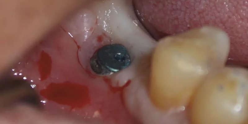
Once all multi-unit abutments are tight, temporary titanium abutments are attached at 10 Ncm, and the transparent surgical guide is used carefully again to note the exact positioning and inform the opening of insertion channels through the interim prosthesis. This must eventually seat freely onto the mucosa, exactly like an immediate denture without interference from metal components once all the channels are completed.
The final position can be checked visually, focusing on features such as an esthetic occlusal plane, incisal display, lip support, centric occlusion, and alignment of center lines or checked using a putty bite provided by the laboratory from the articulated setup.
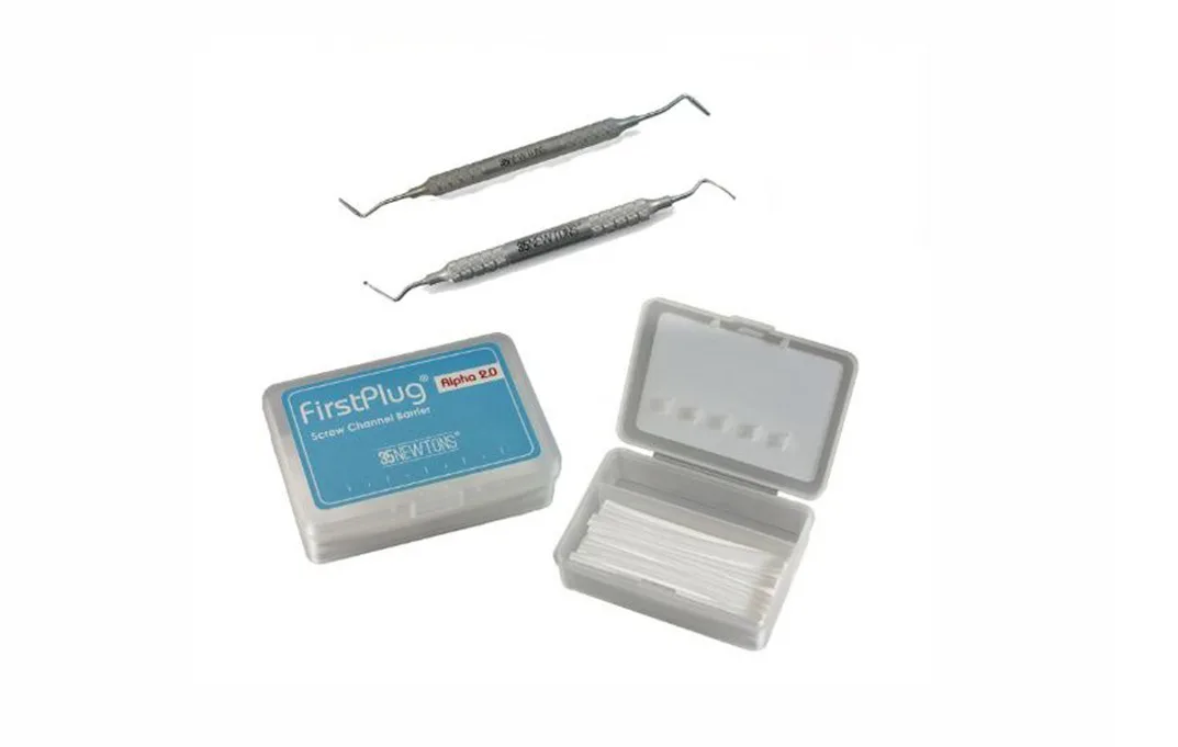
The prosthesis is then washed and dried, and a bonding agent is applied to the channels. The prosthesis is then attached using an intraoral reline process and removed for extraoral final finishing by uncovering and counter rotating the screws, which have been protected by packing PTFE tape to the full height of each hollow abutment (Boulos, 2010).
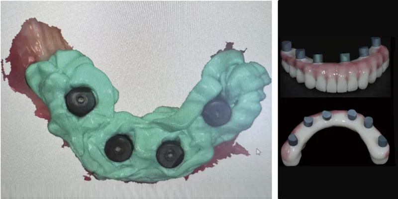
The alternative to picking up all supragingival abutments intraorally is to just pick up the anteriors, take a full arch impression with multi-unit impression abutments, and transfer both to the laboratory to create the posterior access and finish the bridge on the resulting model.
The author finds this alternative keeps the patient in the chair longer, especially if inadvertent occlusal inaccuracies are introduced by the impression and model production — but in cases where the posterior abutments are at very divergent angles, it results in a stronger, neater interim prosthesis.
The resulting interim acrylic bridge is then inserted with the screws at 10 Ncm and the screw access protected with new PTFE tape packed firmly and finished a millimeter below the prosthetic surface and sealed over with color-matched glass ionomer or resin.
At 4 months, the occlusion is recorded. The bridges are removed and copied using putty. All abutment screws are tightened to 20 Ncm, impression copings are placed at 10 Ncm, and accurate open tray impressions are taken. The bridges are replaced at 10 Ncm, and the patient chooses the final shade and characterization for the definitive try-in.
Once the try-in is successful, the definitive model ridges are reduced by 1 millimeter so that controlled compression is achieved at the final fit. Specific oral hygiene instruction is delivered and emphasized with an illustrated leaflet, and all patients are recalled at 1 month for a 15 Ncm retightening of the prosthetic screws, review of oral hygiene, oral health, and esthetic and occlusal outcome. Patient-appropriate hygiene, screw checking, and radiographic reviews are organized, and feedback recorded.
References
- Abi Nader S, Eimar H, Momani M, Shang K, Daniel NG, Tamimi F. Plaque Accumulation Beneath Maxillary All-on-4 Implant-Supported Prostheses. Clin Implant Dent Relat Res. 2015;17(5): 932-937.
- Afrashtehfar KI. The all-on-four concept may be a viable treatment option for edentulous rehabilitation. Evid Based Dent. 2016; 17(2):56-57.
- Agliardi E, Clericò M, Ciancio P, Massironi D. Immediate loading of full-arch fixed prostheses supported by axial and tilted implants for the treatment of edentulous atrophic mandibles. Quintessence Int. 2010;41(4):285-293.
- Babbush CA, Kutsko GT, Brokloff J. The all-on-four immediate function treatment concept with NobelActive implants: a retrospective study. J Oral Implantol. 2011;37(4):431-445.
- Bedrossian E. Rescue implant concept: the expanded use of the zygoma implant in the graftless solutions. Oral Maxillofac Surg Clin North Am. 2011;23(2):257-76
- Bhering CL, Mesquita MF, Kemmoku DT, Noritomi PY, Consani RL, Barão VA. Comparison between all-on-four and all-on-six treatment concepts and framework material on stress distribution in atrophic maxilla: A prototyping guided 3D-FEA study. Mater Sci Eng C Mater Biol Appl. 2016;69:715-725.
- Boulos PJ. Immediate loading of implants and fixed complete dentures: a simplified prosthetic procedure. Gen Dent. 2010;58(5):406-409.
- Bonesvoll P, Olsen I. Influence of teeth, plaque and dentures on the retention of chlorhexidine in the human oral cavity. J Clin Periodontol. 1974;1(4):214-221.
- Brånemark PI, Hansson BO, Adell R, Breine U, Lindström J, Hallén O, Ohman A. Osseointegrated implants in the treatment of the edentulous jaw – experience from a 10-year period. Scand J Plast Reconstr Surg Suppl. 1977;16:1-132.
- Butura CC, Galindo DF, Jensen OT. Mandibular all-on-four therapy using angled implants: a three-year clinical study of 857 implants in 219 jaws. Oral Maxillofac Surg Clin North Am. 2011;55(4):795-211.
- Chan MH, Holmes C. Contemporary “All-on-4” concept. Dent Clin North Am. 2015; 59(2):421-470.
- de Moraes PH, Olate S, Nóbilo Mde A, Asprino L, de Moraes M, Barbosa Jde A. Maxillary “All-On-Four” treatment using zygomatic implants. A mechanical analysis. Rev Stomatol Chir Maxillofac Chir Orale. 2016;117(2):67-71.
- De Rossi M, Santos CM, Migliorança R, Regalo SC. All on Four® fixed implant support rehabilitation: a masticatory function study. Clin Implant Dent Relat Res. 2014;16(4):594-600.
- Drago C. Cantilever Lengths and Anterior-Posterior Spreads of Interim, Acrylic Resin, Full-Arch Screw-Retained Prostheses and Their Relationship to Prosthetic Complications. J Prosthodont. 2016; doi:10.1111/jopr.12426. [Epub ahead of print]
- Faeghi Nejad M, Proussaefs P, Lozada J. Combining guided alveolar ridge reduction and guided implant placement for all-on-4 surgery: A clinical report. J Prosthet Dent. 2016;115(6):662-667.
- Ferreira EJ, Kuabara MR, Gulinelli JL. “All-on-four” concept and immediate loading for simultaneous rehabilitation of the atrophic maxilla and mandible with conventional and zygomatic implants. Br J Oral Maxillofac Surg. 2010;48(3):218-220.
- Francetti L, Agliardi E, Testori T, Romeo D, Taschieri S, Del Fabbro M. Immediate rehabilitation of the mandible with fixed full prosthesis supported by axial and tilted implants: interim results of a single cohort prospective study. Clin Implant Dent Relat Res. 2008;10(4):255-263.
- Holtzclaw D. All-on-4® Implant Treatment: Common Pitfalls and Methods to Overcome Them. Compend Contin Educ Dent. 2016;37(7):458-465;quiz466.
- Jensen OT, Adams MW, Cottam JR, Parel SM, Phillips WR 3rd. The All-on-4 shelf: maxilla. J Oral Maxillofac Surg. 2010;68(10):2520-2527.
- Jensen OT, Adams MW, Cottam JR, Parel SM, Phillips WR 3rd. The all on 4 shelf: mandible. J Oral Maxillofac Surg. 2011;69(1):175-181.
- Jensen O, Cottam J, Adams M, Adams S. Buccal to lingual transalveolar implant placement for all on four immediate function in posterior mandible: report of 10 cases. J Oral Maxillofac Surg. 2011;69(7):1919-1922.
- Jensen OT, Cottam JR, Ringeman JL, Graves S, Beatty L, Adams MW. Angled dental implant placement into the vomer/nasal crest of atrophic maxillae for All-on-Four immediate function: a 2-year clinical study of 100 consecutive patients. Int J Oral Maxillofac Implants. 2014;29(1):e30-e35.
- Khatami AH, Smith CR. “All-on-Four” immediate function concept and clinical report of treatment of an edentulous mandible with a fixed complete denture and milled titanium framework. J Prosthodont. 2008;17(1):47-51.
- Krennmair S, Seemann R, Weinländer M, Krennmair G, Piehslinger E. Immediately loaded distally cantilevered fixed mandibular prostheses supported by four implants placed in both fresh extraction and healed sites: 2-year results from a prospective study. Eur J Oral Implantol. 2014;7(2):173-184.
- Li X, Cao Z, Qiu X, Tang Z, Gong L, Wang D. Does matching relation exist between the length and the tilting angle of terminal implants in the all-on-four protocol? stress distributions by 3D finite element analysis. J Adv Prosthodont. 2015;7(3):240-248.
- Listgarten MA, Lai CH. Ultrastructure of the intact interface between an endosseous epoxy resin dental implant and the host tissues. J Biol Buccale. 1975;3(1):13-28.
- Lopes A, Maló P, de Araújo Nobre M, Sánchez-Fernández E, Gravito I. The NobelGuide® All-on-4® Treatment Concept for Rehabilitation of Edentulous Jaws: A Retrospective Report on the 7-Years Clinical and 5-Years Radiographic Outcomes. Clin Implant Dent Relat Res. 2017;9(3):e474-e488.
- Maló P, de Araújo Nobre M, Lopes A, Moss SM, Molina GJ. A longitudinal study of the survival of All-on-4 implants in the mandible with up to 10 years of follow-up. J Am Dent Assoc. 2011;142(3):310-320.
- Maló P, Nobre Md, Lopes A. Immediate loading of ‘All-on-4’ maxillary prostheses using trans-sinus tilted implants without sinus bone grafting: a retrospective study reporting the 3-year outcome. Eur J Oral Implantol. 2013;6(3):273-283.
- Meloni SM, Tallarico M, Pisano M, Xhanari E, Canullo L. Immediate Loading of Fixed Complete Denture Prosthesis Supported by 4-8 Implants Placed Using Guided Surgery: A 5-Year Prospective Study on 66 Patients with 356 Implants. Clin Implant Dent Relat Res. 2017;19(1):195-206.
- Nazirkar G, Bhanushall S, Singh S, Pattanaik B, Raj N. Effect of Anatase Titanium Dioxide Nanoparticles on the Flexural Strength of Heat Cured Poly Methyl Methacrylate Resins; An In-vitro Study. J Indian Prosthodont Soc. 2014;14(suppl 1):144-149.
- Patzelt SB, Bahat O, Reynolds MA, Strub JR. The all-on-four treatment concept: a systematic review. J Clin Dent Relat Res. 2014;16:836-855.
- Pomares C. A retrospective clinical study of edentulous patients rehabilitated according to the ‘all on four’ or the ‘all on six’ immediate function concept. Eur J Oral Implantol. 2009;2(1): 55-60.
- Saleh Saber F, Ghasemi S, Koodaryan R, Babaloo A, Abolfazli N. The Comparison of Stress Distribution with Different Implant Numbers and Inclination Angles In All-on-Four and Conventional Methods in Maxilla: A Finite Element Analysis. J Dent Res Dent Clin Dent Prospects. 2015;9(4):246-253.
- Silva GC, Mendonça JA, Lopes LR, Landre J Jr. Stress patterns on implants in prostheses supported by four or six implants: a three-dimensional finite element analysis. Int J Oral Maxillofac Implants. 2010;5(2):239-246.
- Tallarico M, Canullo L, Pisano M, Peñarrocha-Oltra D, Peñarrocha-Diago M, Meloni SM. An up to 7-Year Retrospective Analysis of Biologic and Technical Complication With the All-on-4 Concept. J Oral Implantol. 2016;42(3):265-271.
Stay Relevant With Implant Practice US
Join our email list for CE courses and webinars, articles and mores