Educational aims and objectives
This self-instructional course for dentists aims to identify the causes and contributing factors to poor implant esthetics and offers keys for long-term success.
Expected outcomes
Implant Practice US subscribers can answer the CE questions by taking the quiz to earn 2 hours of CE from reading this article. Correctly answering the questions will demonstrate the reader can:
- Identify some factors influencing implant esthetics.
- Recognize how biology influences tissue behavior.
- Realize the role of tissue response to surgery and restorative procedures.
- Identify the five risk factors associated with peri-implant soft tissue dehiscences (PSTDs).
- Observe a case study where the clinician was faced with esthetic challenges.
Dr. David Wong writes about the causes of poor implant esthetics and offers strategies for maintaining long-term esthetics and stability.
Dr. David Wong offers guidance on establishing soft tissue esthetics and stability to implants
Introduction
Replacing missing anterior teeth with dental implants can make a huge positive impact on a patient’s self-image, beauty, and confidence; however, poor implant esthetics can have the opposite effect, leaving patients ashamed to smile. The treatment to recover from poor implant esthetics can be a long, arduous, expensive, and painful process. Often, attempts to correct implant appearances still leave the patient unsatisfied. This article and case study is aimed at identifying the causes and contributing factors to poor implant esthetics and offer keys for long-term success.
5 key points
- Poor implant esthetics are often due to poor gingival form and/or poor tissue quality.
- Implant esthetics are influenced by implant position and depth.
- Common gingival defects surrounding implants include poor gingival levels, lack of interdental papillae, or inadequate soft tissue contour.
- The quality of implant esthetics tends to decrease over
- Surgical techniques are available to help establish and stabilize the peri-implant soft tissue during implant
Abstract
Avoiding poor implant esthetics involves careful evaluation, planning, and execution. Whether an implant is planned before or after a tooth is extracted, the endpoint of any esthetic implant-supported restoration requires proper management of the hard and soft tissues. It is not merely enough to place a dental implant in a restorable position or even to provide a properly contoured restoration with the correct shade and other important elements. The importance of the surrounding soft tissue should be addressed during the course of dental implant therapy as this is the esthetic variable that is most prone to change over time. Esthetic issues such as soft tissue dehiscences, apical migration of the gingival margin, or loss of papillae height are best anticipated and addressed prior to the delivery of the final restoration. This paper will examine some common surgical strategies to establish soft tissue esthetics and stability over time.
Factors influencing implant esthetics
Implant esthetics are influenced by a number of factors:
- implant position
- existing gingival level
- tissue response to surgery and restorative procedures
- management of the restoration with the abutment and crown
- time1
Implant position
A “restoratively driven” approach to implant placement is often preferred for the best esthetic outcome, which allows for proper abutment and crown design. This can be challenging in areas of severe periodontal destruction, tooth position, ridge atrophy, or the patient’s anatomy. Nevertheless, whether an implant is placed in a fresh extraction socket or a healed site, extremely poor positioning cannot be overcome with surgical or restorative procedures.2,3
Existing gingival levels
A dental implant and restoration cannot be esthetically pleasing by itself. The appearance and alignment of the adjacent teeth should complement each other to develop a pleasing smile. Often, orthodontics or more comprehensive restorative care is necessary to develop the symmetry, soft tissue architecture, and uniformity necessary for the desired esthetic outcome.
Tissue response to surgery and restorative procedures
While it may be simple to perform procedures such as extractions, bone grafting, implant surgery, abutment/crown delivery, etc., it is important to remember that the patient’s biology and wound healing will influence the final outcome. It is impossible to entirely predict how patients will respond to surgery, but their clinical presentation provides some clues. For example, the periodontal phenotype and presence of keratinized tissue are two clinical parameters that surgeons can use to predict future tissue behavior.4,5,6,7 The interproximal height of bone is another important observation to make.8
The periodontal phenotype is often described as either thick or thin. To tell the difference, a common technique is transgingival probing4 where a periodontal probe is inserted into the facial sulcus of a tooth. If the probe is visible through the tissue, the phenotype is thin. If it is not visible, it is considered thick. Thin phenotypes are associated with longer, triangular-shaped teeth with a highly scalloped gingival architecture. There tends to be less keratinized tissue and thinner facial bone, leaving teeth prone to bony dehiscences and fenestrations. Thinner phenotypes tend to respond to insult or injury with gingival recession. Thicker phenotypes tend to be associated with shorter, square-shaped teeth, often surrounded by thicker bands of keratinized tissue which is considered more resistant to gingival recession.9 The flatter gingival architecture of thick phenotypes generally makes implant esthetics more straightforward compared to the thin phenotype.10,11,12
Keratinized tissue around implants is also considered an important part of implant esthetics because like thicker periodontal phenotypes, this tissue is less prone to recession. It is also considered helpful for maintaining implant health in regards to decreasing plaque retention, bleeding, and inflammation.13,14 When surveyed, patients tend to prefer the appearance of keratinized tissue versus non-keratinized tissue.15
The interproximal height of bone (IHB),8 which is the interdental bone height on the mesial and distal side of an implant, is critical to esthetics because it is a key determinant of the presence or absence of papillae and the so-called “black triangles.” To maximize the chances of proper papillae formation/preservation, it is recommended that the IHB is 5 mm or less from the interproximal contracts. The keys to maintaining the IHB are conservative extraction, implant, and regenerative procedures when possible and utilizing methods such as orthodontic extrusion whenever appropriate.
Prosthetic design
Soft tissue esthetics around implants are influenced by the abutment and crown design. Compared to tooth-supported crowns, implant-supported crowns tend to have a flatter or concave subgingival profile16,17 to allow for thicker facial tissue and more stable gingival levels.
Implant esthetics over time
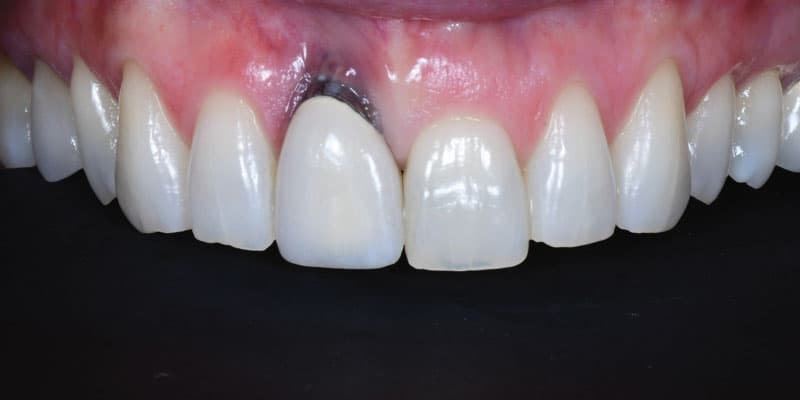
The facial soft tissue changes around implants in the esthetic zone are being studied more as there is now ample data to compare various treatment strategies observed over time. The most common change observed with implants over time is the presence of gingival recession (Figure 1), now called “peri-implant soft tissue dehiscences (PSTDs).”18 In 2000, Small, et al., reported after a 1-year longitudinal prospective study that 80% of implants exhibited approximately 1 mm of recession after the abutment was connected to the implant.19 A study by Cardaropoli, et al., in 2006 found a similar result with the average recession being 0.6 mm at 1 year.20 More recently, Tavelli, et al., in 2022 reported on 176 implants, and 56.8% of them had recession, and 43.2% did not have any noticeable recession.
There were five risk factors associated with PSTDs:
- Time in function
- Implants next to each other
- Bony dehiscence
- Thin facial tissue
- Less keratinized mucosa width.18
Finally, when comparing gingival recession on implants placed in fresh extraction sockets (immediate placement) versus implants placed in a delayed approach, Parvini, et al. (2022), reported increased recession on the immediately placed implants after 12 months (0.37 mm).21 Meanwhile, the delayed placement group exhibited a facial tissue gain of 0.83 mm. Immediate implant placement typically shows the greatest variability in the occurrence of recession with reports ranging from 9% to 41% having at least 1 mm of recession, 1 to 3 years after placement.22,23
Implications for implant treatment
Given the knowledge available regarding soft tissue changes around implants, an increased focus is being placed on the preservation and protection of the facial plate24,25,26,27 improving facial tissue thickness when necessary (i.e., thin phenotypes)7, and the timing of implant placement (immediate versus delayed; serial extraction).11,28 Below is a case study detailing the various techniques available with this hard and soft tissue development philosophy in mind.

Case Study
This 24-year-old white male patient initially presented for an implant consultation to replace his upper central incisors, tooth Nos. 8 and 9. Both teeth had a history of trauma and subsequent root canal therapy; however, No. 8 was recently extracted after a second traumatic event, and No. 9 was deemed endodontically hopeless (Figures 2A and 2B).

This case posed several challenges. Esthetically, the patient had malpositioned teeth and uneven gingival levels. His periodontal phenotype was thin, and he had adequate keratinized mucosa present in the maxilla but much less in the mandible. When considering implant esthetics, specifically, labial ridge atrophy was observed in the No. 8 site (Figure 2C); fortunately, the ridge width was still adequate for proper implant placement (Figures 2D). The other implant challenge was addressing hopeless tooth No. 9. With the thin phenotype and long, tapered tooth morphology, papillae preservation and the stability of the facial tissue was a major concern. There was no loss of the interproximal height of bone, so with tooth No. 9 still present, the proper surgical strategy and timing was crucial to ensure preservation of the tissues. The final challenge was patient management. He self-reported that he is “rough with his teeth” which has resulted in his present situation.
Implant esthetics is influenced by four main factors:
- existing gingival levels
- implant position
- response of the tissues to dental/surgical procedures
- management of the tissue with the crown/abutment1
The patient agreed that orthodontics and two implants was the ultimate goal.
Considering the esthetic risk of two implants side-by-side, a serial extraction (aka as “one by one”) approach,11,28 was taken in order to preserve the midline papilla and the interdental bone between the implants. In this case, tooth No. 8 was treated with an implant first, followed by extraction and treatment of tooth No. 9 approximately 3 months later. Both implants were placed in positions to both accommodate screw-retained restorations as well as to allow thicker facial bone to avoid future bony dehiscences (more lingual position).29
To counteract the tendency of anterior implants to develop soft tissue dehiscences (PSTDs), each implant site was managed differently, since tooth No. 8 was a healed site, and tooth No. 9 was still present. Tooth No. 8 was treated with an implant and a simultaneous connective tissue graft to address the facial ridge atrophy.30 Tooth No. 9 was treated with the “socket shield” technique, which is a form of root submergence where a thin facial portion of the root (the root shield) is left attached to the facial plate in order to avoid any loss of facial bone and tissue.


The sequence of treatment was the following:
- Referred to orthodontist for evaluation and treatment prior to commencement of any implant-related surgery (Figure 2E).
- Placed implant No. 8 with the addition of a connective tissue graft and healing abutment (Figures 2F-2H). With this technique, a soft tissue allograft such as an acellular dermal graft material may also be used.3
- After 3 months, removed tooth No. 9 utilizing the “socket shield”25,26,27 approach and grafted the socket (Figures 2I-2K).
- After 3 months of healing of the grafted socket of tooth No. 9, a surgical guide (which was also used for tooth No. 8) was used to place implant No. 9 (Figures 2L and 2M).
- Implant No. 9 was allowed to heal for 4 months, and both sites Nos. 8 and 9 were evaluated (Figures 2N-2P). The facial gingival thickness and keratinized tissues were satisfactory; however, recession and a mucogingival deficiency had developed on tooth Nos. 24-25 post-orthodontically.
- The gingival levels between tooth Nos. 8-9 were uneven; this was corrected through the provisional restorations (Figures 2Q and 2R).
- After addressing the recession on tooth Nos. 24-25 with a free gingival graft, final restorations were placed on tooth Nos. 8 and 9, making desired changes from the patient (Figures 2S and 2T). Figure 2T is 4 years posttreatment.


Conclusions
Implant esthetics requires a lot of consideration and planning to achieve the desired result. While there are many approaches available, especially in the case presented, it is important to understand how biology influences tissue behavior. The present focus is on maximizing tissue thickness, minimizing bone loss after extraction, placing implants away from the facial plate, and tissue management using the abutments and crowns. Other surgical strategies such as immediate implant placement or perhaps even a single implant to replace both teeth8 are other options that were considered. No matter the treatment plan, one must always treat to their ability and take advantage of interdisciplinary approaches such orthodontics, periodontics, and restorative dentistry (in this case) whenever appropriate.
Dr. Bernard Touati offers his insights into effective anterior implant esthetics. Read his article here: https://implantpracticeus.com/anterior-implant-esthetics/
Author Info
 David Wong, DDS, is a board-certified periodontist in fulltime private practice in Tulsa, Oklahoma. He received his undergraduate education and dental training at the University of Oklahoma. He then went on to complete a 3-year residency in periodontics at the University of Missouri-Kansas City. He is a Diplomate of the American Board of Periodontology and holds teaching positions at the University of Oklahoma College of Dentistry and Penn Dental Medicine Periodontic Program. He is a published author in several peer-reviewed dental journals and has also reached a mainstream audience in media such as Fox News and the Wall Street Journal. Dr. Wong presently resides in Tulsa with his wife and three children.
References
- Cooper LF. Objective criteria: guiding and evaluating dental implant esthetics. J Esthet Restor Dent. 2008;20(3):195-205.
- Nisapakultorn K, Suphanantachat S, Silkosessak O, Rattanamongkolgul S. Factors affecting soft tissue level around anterior maxillary single-tooth implants. Clin Oral Implants Res. 2010 Jun;21(6):662-670.
- Park JB. Increasing the width of keratinized mucosa around endosseous implant using acellular dermal matrix allograft. Implant Dent. 2006 Sep;15(3):275-281.
- Fu J, Lee A, Wang HL (2011) Influence of tissue biotype on implant esthetics. Int J Oral Maxillofac Implants. 26:499–508
- Fu JH, Su CY, Wang HL. Esthetic soft tissue management for teeth and implants. J Evid Based Dent Pract. 2012 Sep;12(3 Suppl):129-142.
- Jung RE, Sailer I, Hämmerle CH, Attin T, Schmidlin P. In vitro color changes of soft tissues caused by restorative materials. Int J Periodontics Restorative Dent. 2007 Jun;27(3):251-257.
- Kan JYK, Yin S, Rungcharassaeng K, Zucchelli G, Urban I, Lozada J. Facial implant gingival level and thickness changes following maxillary anterior immediate tooth replacement with scarf-connective tissue graft: A 4-13-year retrospective study. J Esthet Restor Dent. 2023 Jan;35(1):138-147.
- Salama H, Salama MA, Garber D, Adar P. The interproximal height of bone: a guidepost to predictable aesthetic strategies and soft tissue contours in anterior tooth replacement. Pract Periodontics Aesthet Dent. 1998 Nov-Dec;10(9):1131-1141; quiz 1142.
- Magne P, Belser U. Bonded porcelain restorations in the anterior dentition. A biomimetic approach. Chicago: Quintessence; 2002.
- Kao RT, Fagan MC, Conte GJ. Thick vs. thin gingival biotypes: a key determinant in treatment planning for dental implants. J Calif Dent Assoc. 2008 Mar;36(3):193-198.
- Khoury G, Chamieh F, Fromentin O. One-by-one immediate dental implants: A papillae preservation concept for adjacent implants in a compromised periodontal case. Clin Case Rep. 2020 Aug 20;8(12):2664-2672.
- Kois JC, Kan JY. Predictable peri-implant gingival aesthetics: surgical and prosthodontic rationales. Pract Proced Aesthet Dent. 2001 Nov-Dec;13(9):691-698; quiz 700, 721-722.
- Chung DM, Oh TJ, Shotwell JL, Misch CE, Wang HL. Significance of keratinized mucosa in maintenance of dental implants with different surfaces. J Periodontol. 2006 Aug;77(8):1410-1420.
- Warrer K, Buser D, Lang NP, Karring T. Plaque-induced peri-implantitis in the presence or absence of keratinized mucosa. An experimental study in monkeys. Clin Oral Implants Res. 1995 Sep;6(3):131-138.
- Bonino F, Steffensen B, Natto Z, Hur Y, Holtzman LP, Weber HP. Prospective study of the impact of peri-implant soft tissue properties on patient-reported and clinically assessed outcomes. J Periodontol. 2018 Sep;89(9):1025-1032.
- Gomez-Meda R, Esquivel J, Blatz MB. The esthetic biological contour concept for implant restoration emergence profile design. J Esthet Restor Dent. 2021 Jan;33(1): 173-184.
- Rompen E, Raepsaet N, Domken O, Touati B, Van Dooren E. Soft tissue stability at the facial aspect of gingivally converging abutments in the esthetic zone: a pilot clinical study. J Prosthet Dent. 2007 Jun;97(6 Suppl):S119-125.
- Tavelli L, Barootchi S, Majzoub J, Chan HL, Stefanini M, Zucchelli G, Kripfgans OD, Wang HL, Urban IA. Prevalence and risk indicators of midfacial peri-implant soft tissue dehiscence at single site in the esthetic zone: A cross-sectional clinical and ultrasonographic study. J Periodontol. 2022 Jun;93(6):857-866.
- Small PN, Tarnow DP. Gingival recession around implants: a 1-year longitudinal prospective study. Int J Oral Maxillofac Implants. 2000 Jul-Aug;15(4):527-532.
- CCardaropoli G, Lekholm U, Wennström JL. Tissue alterations at implant-supported single-tooth replacements: a 1-year prospective clinical study. Clin Oral Implants Res. 2006 Apr;17(2):165-171.
- Parvini P, Müller KM, Cafferata EA, Schwarz F, Obreja K. Immediate versus delayed implant placement in the esthetic zone: a prospective 3D volumetric assessment of peri-implant tissue stability. Int J Implant Dent. 2022 Nov 25;8(1):58.
- Chen ST, Buser D. Clinical and esthetic outcomes of implants placed in postextraction sites. Int J Oral Maxillofac Implants. 2009;24 Suppl:186-217.
- Chen ST, Buser D. Esthetic outcomes following immediate and early implant placement in the anterior maxilla–a systematic review. Int J Oral Maxillofac Implants. 2014;29 Suppl:186-215.
- Agnini A, Salama MA, Salama H, Garber D, Agnini AM. Surgical veneer grafting: compensation for natural labial plate remodeling after immediate implant placement. J Cosmetic Dent. 2017 Winter; 32(4):70-85.
- Gluckman H, Du Toit J, Salama M. The Pontic-Shield: Partial Extraction Therapy for Ridge Preservation and Pontic Site Development. Int J Periodontics Restorative Dent. 2016 May-Jun;36(3):417-423.
- Gluckman H, Salama M, Du Toit J. Partial Extraction Therapies (PET) Part 1: Maintaining Alveolar Ridge Contour at Pontic and Immediate Implant Sites. Int J Periodontics Restorative Dent. 2016 Sep-Oct;36(5):681-687.
- Gluckman H, Du Toit J, Salama M, Nagy K, Dard M. A decade of the socket-shield technique: a step-by-step partial extraction therapy protocol. Int J Esthet Dent. 2020;15(2):212-225.
- Funato A, Salama MA, Ishikawa T, Garber DA, Salama H. Timing, positioning, and sequential staging in esthetic implant therapy: a four-dimensional perspective. Int J Periodontics Restorative Dent. 2007 Aug;27(4):313-323.
- Rojas-Vizcaya F. Biological aspects as a rule for single implant placement. The 3A-2B rule: a clinical report. J Prosthodont. 2013 Oct;22(7):575-580.
- Seibert JS, Salama H. Alveolar ridge preservation and reconstruction. Periodontol 2000. 1996 Jun;11:69-84.
Stay Relevant With Implant Practice US
Join our email list for CE courses and webinars, articles and mores

