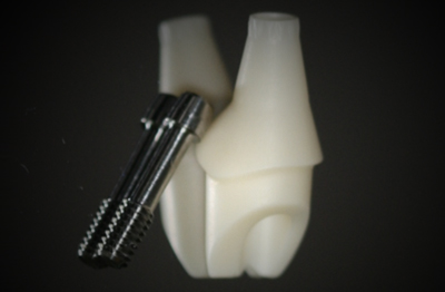
Drs. John Lupovici and Robert Raimondi illustrate a collaborative surgical and restorative clinical treatment for esthetic replacement of a compromised maxilla central incisor
With the rapid advancement in understanding of the biomechanical interaction of implants and bone, implants no longer can be deemed successful simply based on their ability to retain a functional restoration.
[userloggedin]
Instead, the faithful re-creation of natural esthetics has also become imperative. In order to solve the substantial challenge of achieving natural esthetics in compromised situations, a number of techniques and materials have developed to restore deficient alveolar bone and maintain existing bone and soft tissue. This case study will exhibit an example of a collaborative surgical and restorative clinical treatment to achieve an esthetic replacement of a compromised maxilla central incisor.
Case report
The 31-year-old male patient presented with asymptomatic external resorption of the right central incisor. A review of his medical and social history proved uneventful. Extraoral examination revealed a moderate to high smile line that displayed approximately 2 mm of gingiva when he smiled. Intraoral and radiographic examination revealed the presence of external palatal resorption of the maxillary right central incisor, in conjunction with a thin gingival biotype (Figures 1 and 2).
Although a variety of bone-augmentation strategies (onlay, inlay, veneer grafts, ridge splitting, and expansion) are all well documented to have a record rate of success, we chose a staged extraction and guided bone regeneration (GBR) technique because of the patient’s prominent smile line in conjunction with thin gingival biotype (Figure 1).
Following administration of local anesthesia (Lidocaine with epinephrine 1:100,000), tooth No. 8 was atraumatically elevated from the alveolus, and socket augmentation was performed with the particulate mineralized allograft (Musculoskeletal Transplant Foundation Symbios™ Mineralized Cortical/Cancellous Granules, .5 cc (Dentsply Implants) (Figure 3) and a collagen sponge. The patient was provided with a non-load-bearing Essix retainer. Three months after the extraction and augmentation, implant placement using OsseoSpeed™ (4.5 x 13 mm) (Dentsply Astra Tech) implants was performed.
Six weeks after implant placement, implant integration was confirmed (Figure 6). An open-tray impression was made using Impregum™ soft medium body (3M™ ESPE™) in a custom tray. Using the patient’s original tooth, a zirconia custom Atlantis™ abutment (Atlantis Abutments, Dentsply Implants) was reverse engineered (Figure 8). The abutment (Figure 9) was then delivered and torqued to 25 Ncm, and a provisional restoration was fabricated (Figure 10). Over the course of 3 months, the tissue was matured prior to fabrication of the final restoration. The final restoration was then delivered successfully (Figure 11).
Gallery
{gallery}140310_cl_lupovici{/gallery}
Stay Relevant With Implant Practice US
Join our email list for CE courses and webinars, articles and mores


