Dr. Ara Nazarian discusses materials and methods that can help fulfill patients’ surgical and restorative needs
When a patient presents to your dental practice with questionable and/or non-restorable teeth requiring full mouth extractions, the biggest concern is whether or not implants can be placed at the same surgical visit and, if so, if the patient will be able to walk out with fixed teeth. Having the ability to place implants within your practice allows you to load or progressively load, so you can meet the needs of these particular patients; this lets you position your practice on a whole new level. Of course, certain parameters must be met in order to facilitate this type of treatment. This includes, but is not limited to, the quality and quantity of bone, the presence of infection, the patient’s health, and the skills of the dental provider. Additionally, the selection of the most appropriate materials for the most ideal situation must be met.
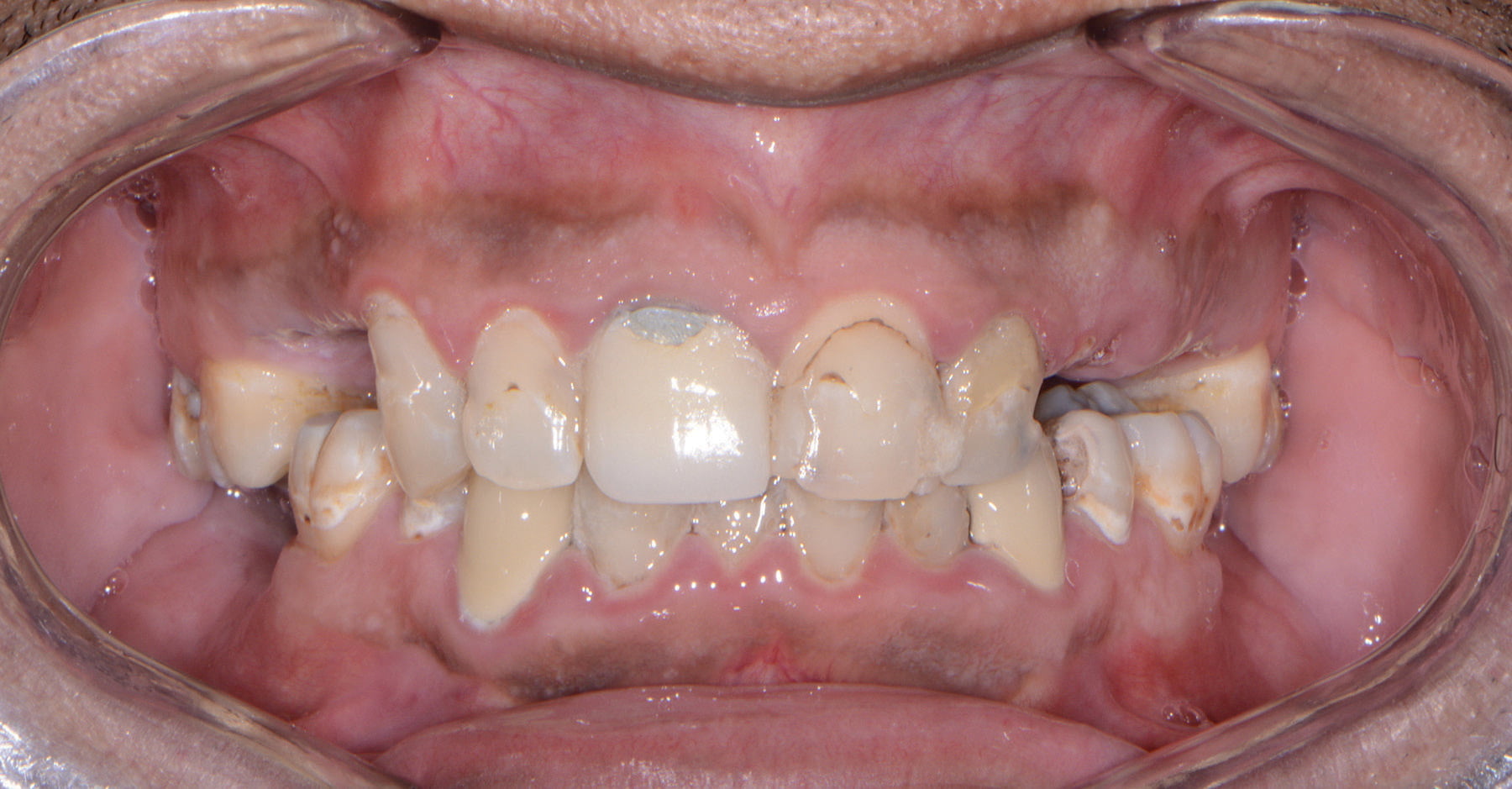
Figure 1: Preoperative retracted frontal view


Figure 2: Preoperative maxillary occlusal view and Figure 3: Preoperative mandibular occlusal view
A patient presented to my practice for a consultation wanting to restore his dentition to proper form and function (Figure 1). He complained of generalized discomfort in these teeth due to the gross caries and perio-dontal disease that were readily apparent (Figures 2 and 3). There were several teeth in both arches, which had so much extensive decay, that only the root tips were apparent upon clinical examination. Also, there was hyper-eruption in certain areas of his posterior dentition, as well as a deep impinging bite in the anterior.
Planning
The clinical evaluation included information regarding lip length and support, existing tooth position of the natural teeth, occlusion, restorative space, and phonetics. In addition, digital images of frontal, side, and occlusal views of the dentition as well as facial shots were captured with a Nikon D7200 (Photo Med).
A CBCT scan and panoramic radiograph were taken using the CS 8100 3D (Carestream Dental) (Figures 4 and 5) to accurately capture the information needed to properly treatment plan this case ensuring the most ideal outcome, especially since the patient discussed his frustration with previous treatment that did not last very long or address his primary needs or requests. Using the CS 3D Imaging software (Carestream Dental), dental implants were virtually planned in key positions in both maxillary and mandibular arches (Figure 6).
To further develop a treatment plan, diagnostic model impressions were taken using Panasil® (Kettenbach) heavy and light body polyvinyl siloxane impression material (Figures 7 and 8), poured up and forwarded to the dental lab. These models were then mounted on an articulator (Stratos® 100, Ivoclar Vivadent) for further analysis in order to meet the patient’s esthetic and functional needs.
Financing options using a third-party payment option (Lending Club) were discussed with the patient. This discussion was a very important part of facilitating acceptance of his care, since it made the cost of treatment more feasible.
A 3D virtual treatment plan was further developed from our planning with the CS 3D imaging software and integrating with the photos and models with the assistance of 3DDX (Figure 9). A virtual online integrative meeting with 3DDX allowed for a comprehensive review of the assembled digital and clinical information formulating an optimal treatment plan that would fulfill the necessary requirements for esthetics, form, and function.

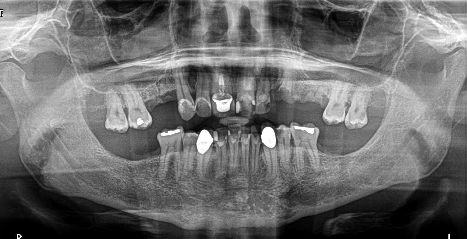
Figure 4: CS 8100 3D and Figure 5: Preoperative panoramic image
The implants that would be utilized for this case were OCO Biomedical’s Engage™ dental implants. These implants are known for their unchallenged high implant stability at placement, which is a critical success factor in these immediate load cases. With the combination of their patented Bull Nose Auger™ tip and Mini Cortic-O Thread™, the Engage™ implant system offers practitioners a bone level implant with high initial stability for selective loading options.
The Engage™ implant is self-tapping for an enhanced mechanical lock in the bone. The Bull Nose Auger™ tip will not proceed any deeper than the initial pilot drill preparation locking into the base of the osteotomy. Engage™ implants have a proprietary surface treatment designed to increase the surface area of the implant for optimal bone in-growth and stability.
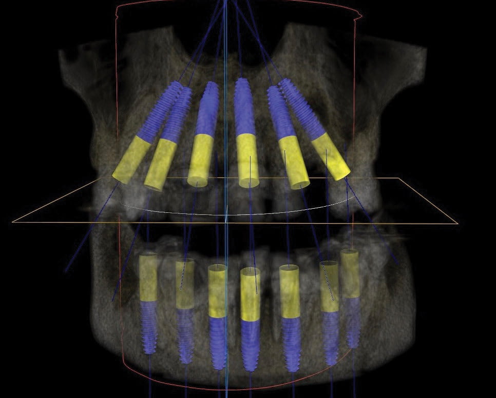

Figure 6: Proposed treatment planned in CS 3D Imaging software and Figure 7: Maxillary impression (Kettenbach)
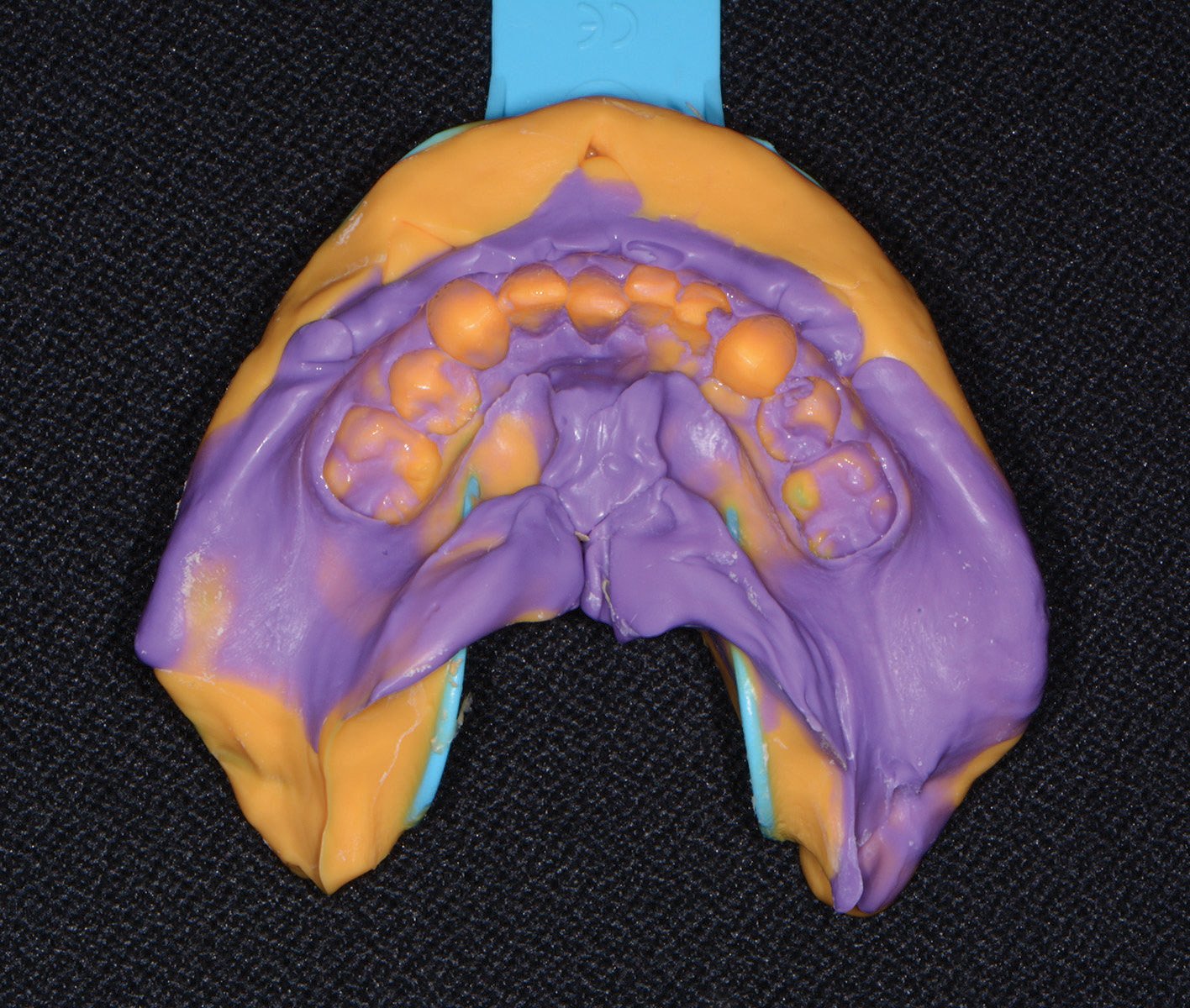
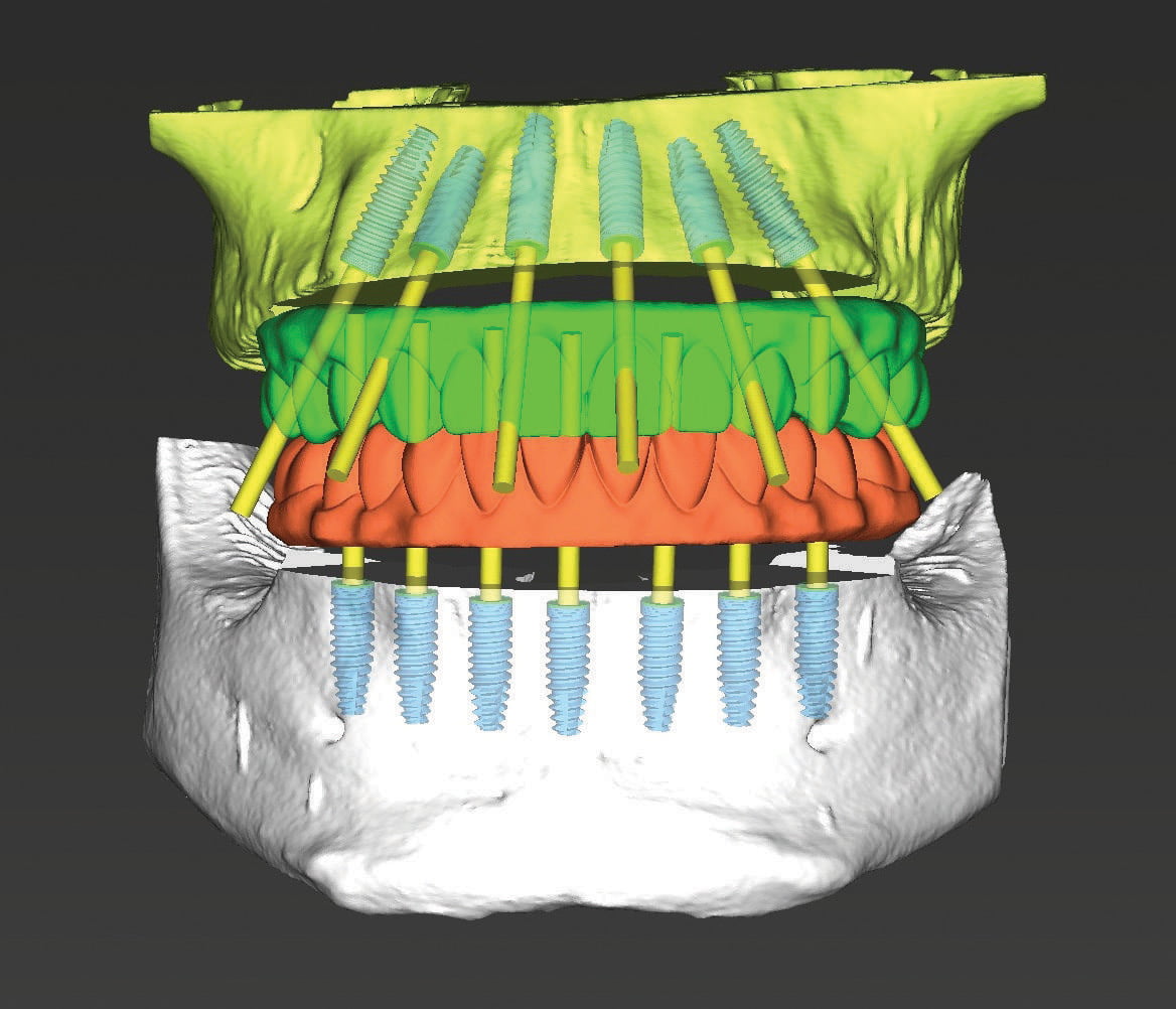
Figure 8: Mandibular impression (Kettenbach) and Figure 9: 3DDX virtual treatment plan maxilla/mandible
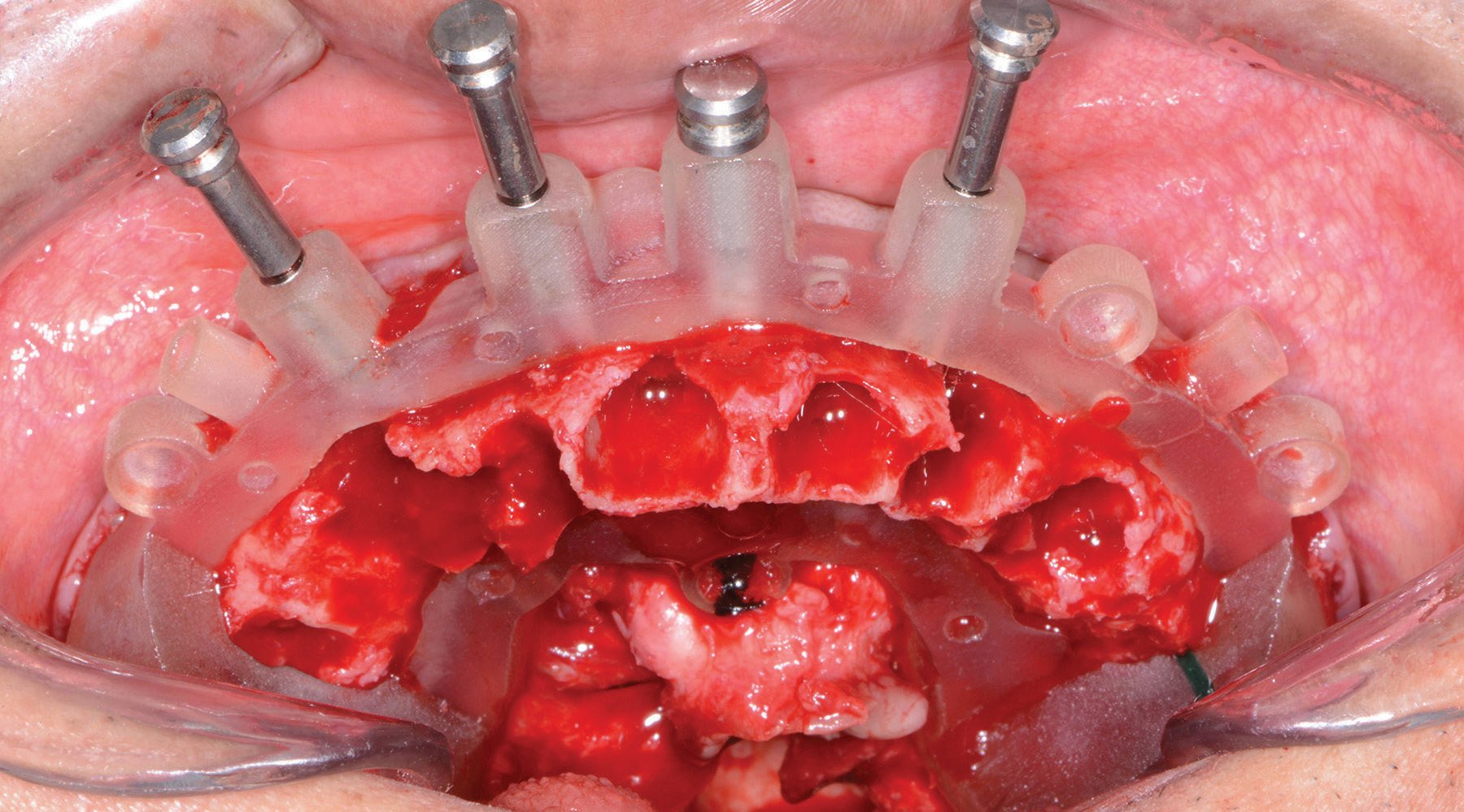
Figure 10: Maxillary bone leveling guide
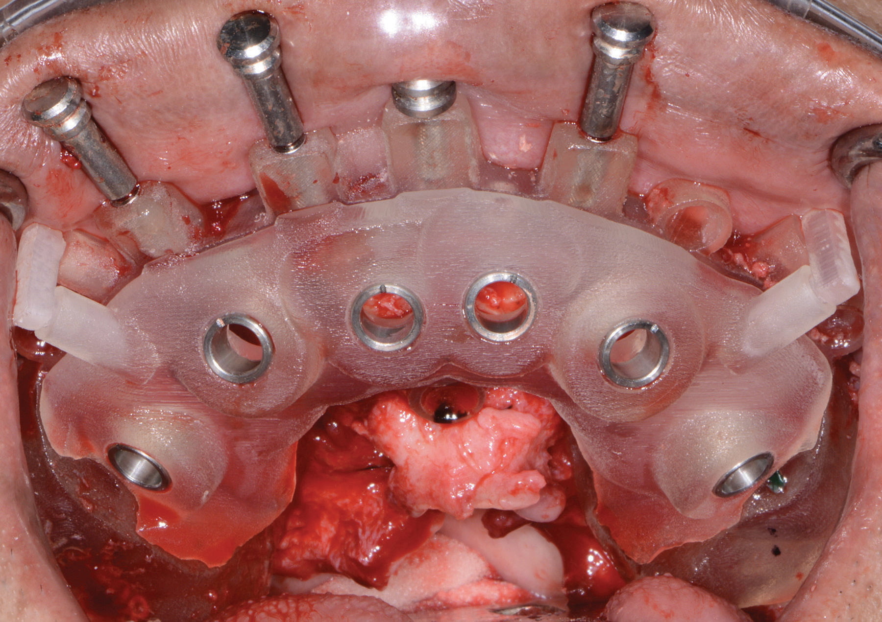
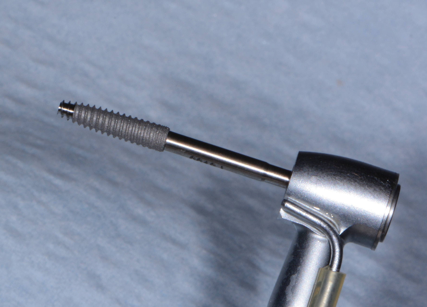
Figure 11: Maxillary implant surgical guide and Figure 12: Engage (OCO Biomedical) dental implant
Once the virtual plan was orchestrated and fully confirmed, the next appointment would be the planned surgery. The patient was appropriately sedated with IV medications, and local anesthesia was administered in both arches. The maxillary teeth were atraumatically extracted utilizing the Physics Forceps™ (GoldenDent). The tissue was then reflected using the Reflector (GoldenDent) instrument, so that the bone leveling surgical guide (3DDX) would be fully seated and fixed with its respectful retention pins (Figure 10).

Figure 13: Implants with corresponding multiunit abutments
Once the appropriate bone leveling was accomplished with the surgical handpiece, the universal implant surgical guide was positioned into the bone leveling guide, and the sites for the implants were initiated with a designated 1.8 mm pilot drill in its appropriate key from the OCO Biomedical Guided Kit (Figure 11) utilizing the Mont Blanc surgical handpiece and Aseptico® surgical motor (AEU 7000) at a speed of 1200 rpm with copious amounts of sterile saline. Sequential osteotomy formers and keys from the OCO Biomedical Guided Kit were then used to shape the final osteotomies. Once the osteotomies were complete, a rotary implant driver was used to place the dental implants until increased torque was necessary (Figure 12). The ratchet wrench was then connected to the adapter, and the implants torqued to final depths reaching a torque level of about 40-50Ncm.
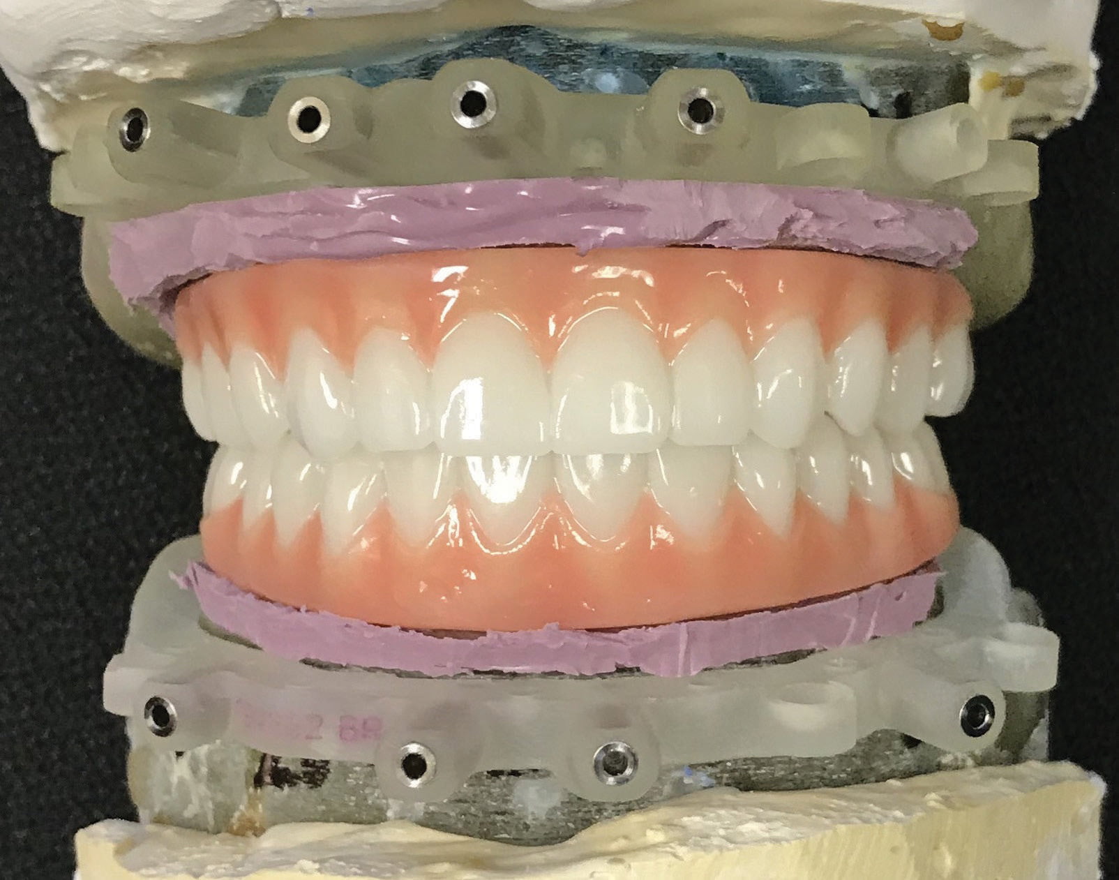
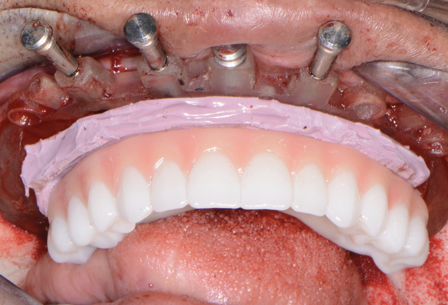
Figure 14: PMMA maxillary/mandibular restorations and Figure 15: Insertion of provisional restoration
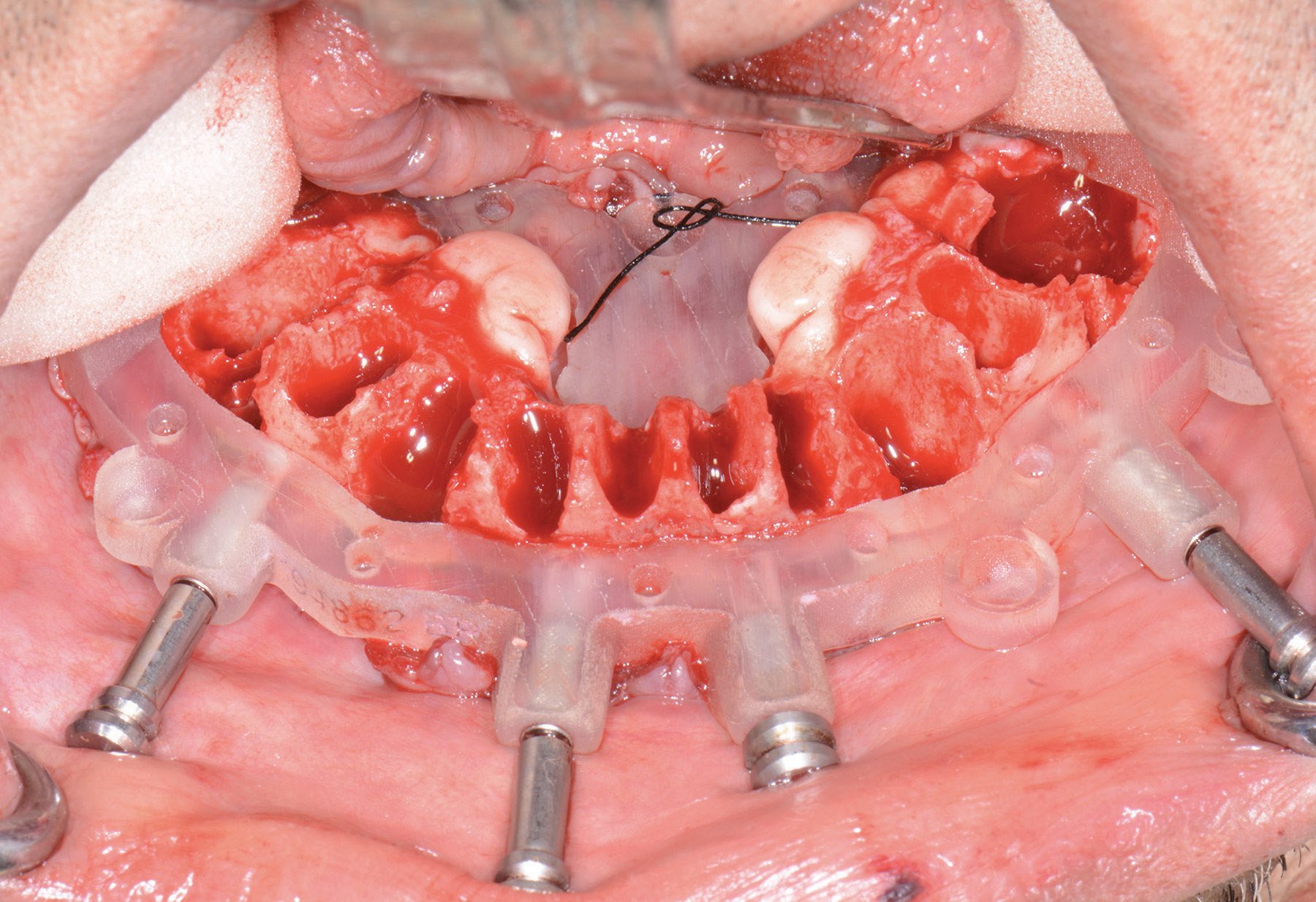
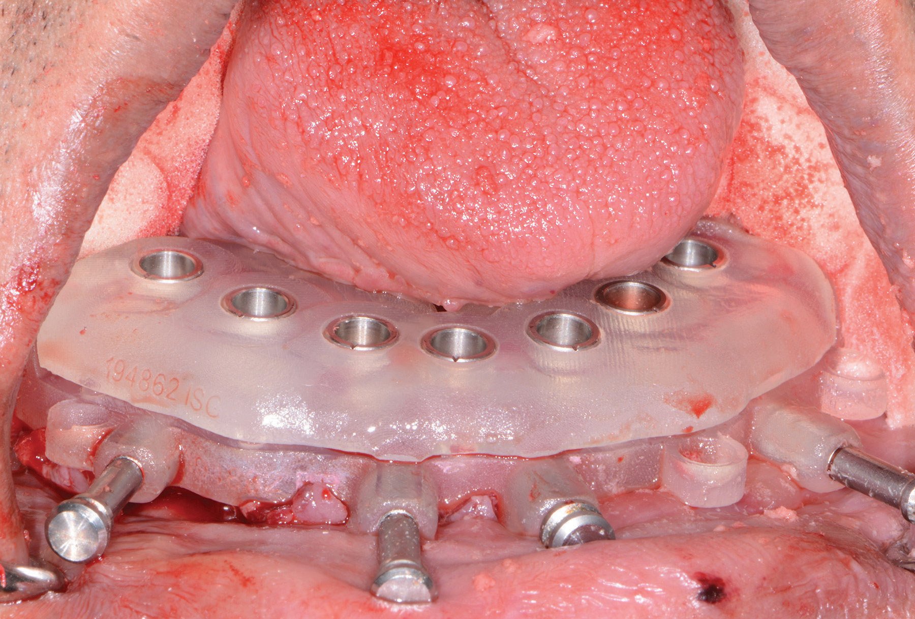
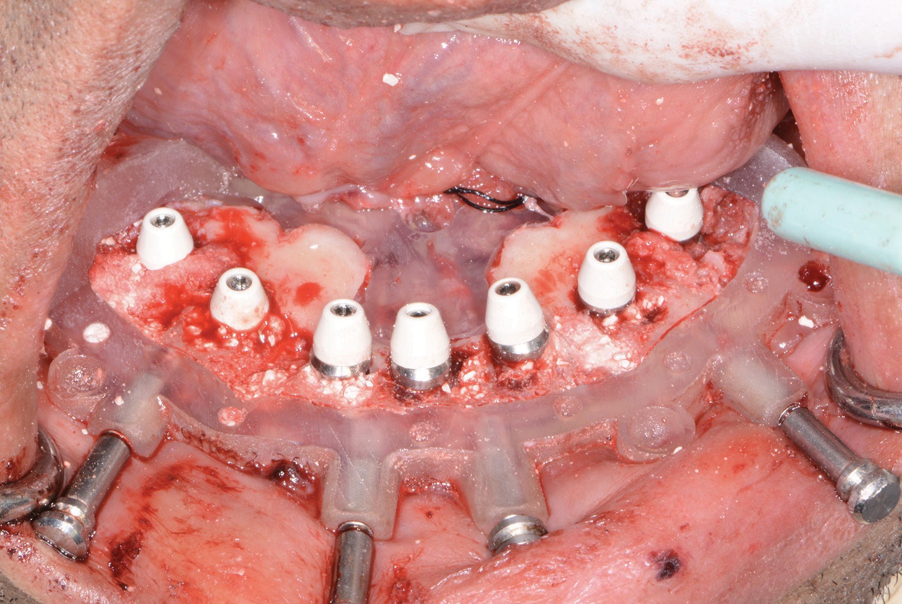
Figure 16: Mandibular bone leveling guide and Figure 17: Mandibular implant surgical guide and Figure 18: Implants with corresponding multiunit abutments
A baseline ISQ reading was taken of these implants utilizing the Penguin (Aseptico) RFA unit. Since the initial readings were all above 70 and the quality of bone after leveling was good, multiunit abutments (OCO Biomedical) were tightened into the Engage (OCO Biomedical) dental implants at 25Ncm followed by temporary cylinders at 15Ncm.
Any residual areas around the implants or in the sockets were grafted with a cortical mineralized and demineralized bone grafting material to optimize the area for regeneration (Figure 13).
The prefabricated immediate provisional arch restorations (3DDX) with pre-drilled access openings were inspected before being tried in (Figure 14).
The maxillary provisional restoration was tried in to verify a passive fit over the temporary abutments. Once confirmed, a polyvinyl siloxane gasket was placed to avoid the restoration (Figure 15) from locking on during the relining procedure with REBASE II Fast Set (Tokuyama®) hard reline material. After the material polymerized, the immediate provisional restoration was removed and any access material removed with the Torque Plus (Aseptico) lab handpiece and acrylic bur (Komet). The same procedures were accomplished in the lower arch (Figures 16, 17, 18); however, while the provisional restoration was being trimmed and polished, the mandibular tori were surgically removed before suturing the soft tissue.
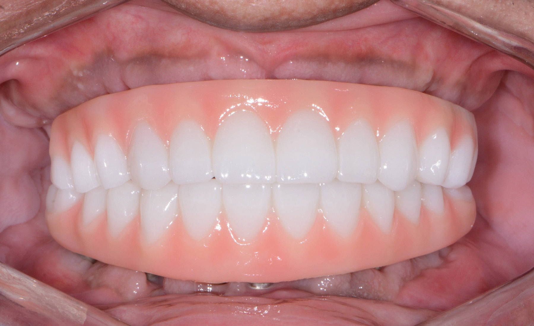
Figure 19: Maxillary and mandibular fixed provisional restorations
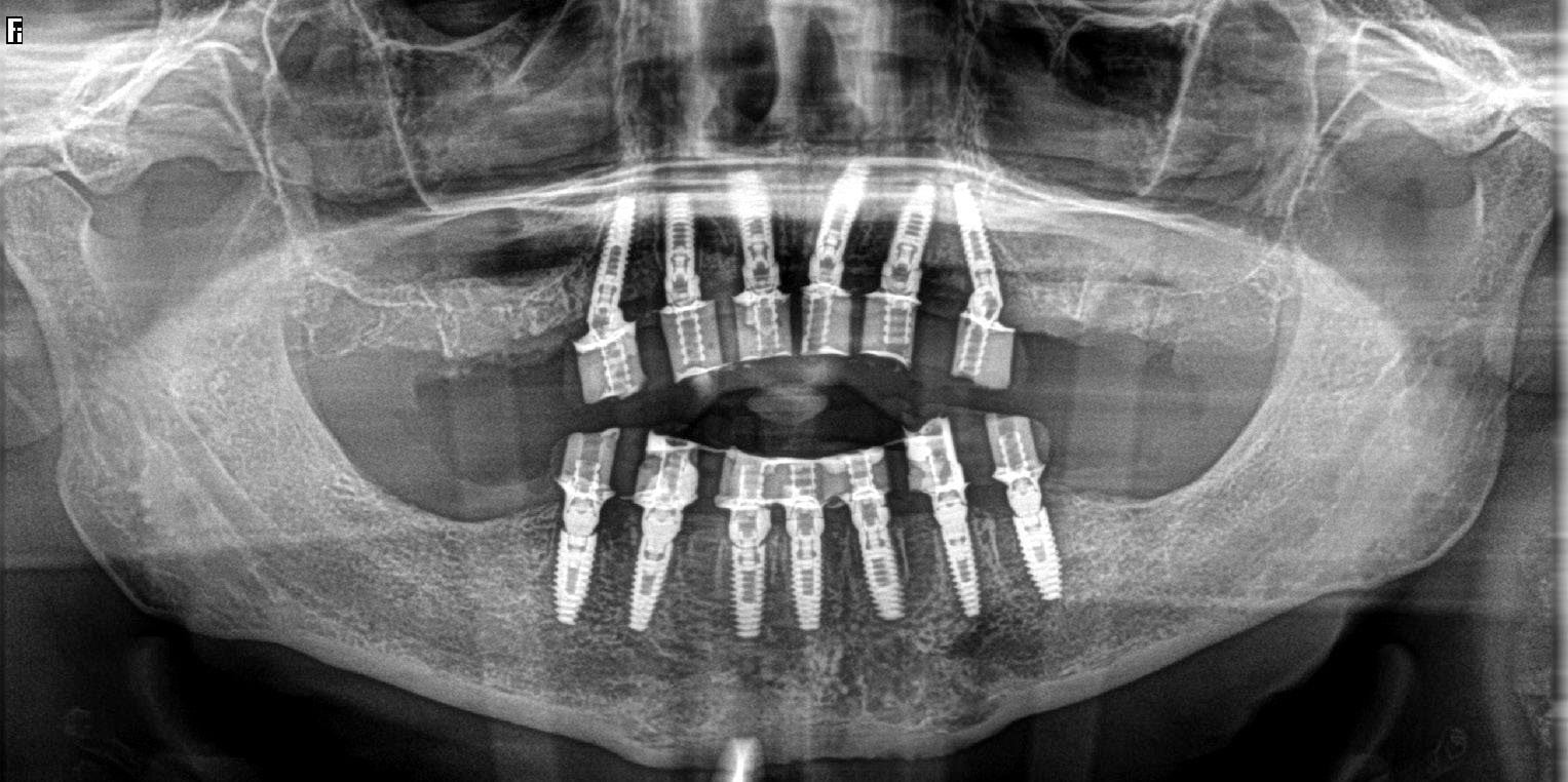
Figure 20: Postoperative panoramic image
Once trimmed and polished, the provisional arch restoration was seated and tightened with a torque wrench at 15Ncm. The access openings were filled three-quarters of the way with Teflon tape followed by Cavit™ (3M) filling material.
Seven days postoperatively, the patient returned with very little discomfort, swelling, or bruising. He was very pleased with his new upper and lower fixed provisional restorations (Figures 19 and 20). Now that the patient was no longer anesthetized, the occlusion was checked again to confirm there were no interferences in lateral and protrusive movements. The next step in his treatment will consist of full arch impressions for the definitive restorations approximately 4 to 5 months postoperatively.
Conclusion
Having the ability to take a patient from start to finish with fewer appointments within your practice allows you to position yourself as a provider that can fulfill your patients’ surgical and restorative needs. With the proper training and appropriate materials, a dental provider may provide extraction, grafting, and implant placement within one appointment at one location. Not only does this allow you to reduce the amount of visits for the patient, but this type of service also helps maintain the cost to the patients since they are not seeing multiple dental providers. Most importantly, this enables the dental provider full control of the surgical and prosthetic outcome. Depending on the patient’s desires, the clinical conditions of the oral environment present, and the skills of the provider, a dentist may choose to extract teeth, level bone, and graft with guided dental implant placement within his/her dental practice.
 Ara Nazarian, DDS, DICOI, maintains a private practice in Troy, Michigan, with an emphasis on comprehensive and restorative care. He is a Diplomate in the International Congress of Oral Implantologists (ICOI) and the director of the Ascend Dental Academy. He has conducted lectures and hands-on workshops on esthetic materials, grafting, and dental implants throughout the United States, Europe, New Zealand, and Australia.
Disclosure: Dr. Nazarian is a Key Opinion Leader for Carestream Dental.
Stay Relevant With Implant Practice US
Join our email list for CE courses and webinars, articles and mores

