Implant Practice US provides continuing education and other valuable resources to help you build upon your expertise in implantology. Our continuing education is updated regularly and accessible any time, from anywhere.
Purchase a subscription today!

Implant treatment in the esthetic zone

Continuing Education (CE)

The continuing education article below is available to Implantologists and general dental practitioners who perform implants.

In order to earn continuing education credits with our publication, you must be a paid subscriber of Implant Practice US and complete a short quiz about the content of the article.

Earn up to 16 online dental CE credits per year! Purchase a subscription now.

Dr. Shakeel Shahdad presents a clinical case illustrating replacement of a maxillary canine with a narrow diameter implant in a hypodontia patient

Implants are now considered a predictable choice to replace missing teeth; nevertheless, replacement in the esthetic zone remains a challenge.

In addition to the challenges posed by the various clinical scenarios, our patients have high expectations; they demand optimal functional and esthetic outcomes.

Educational aims and objectives
This article aims to discuss an implant method of replacing a tooth in the esthetic zone.

Expected outcomes
Implant Practice US subscribers can answer the CE questions to earn 2 hours of CE from reading this article. Correctly answering the questions will  demonstrate the reader can:

  • Identify a method of preoperative assessment for implant placement.
  • Recognize a method to deliver a restoration that esthetically fulfills the objectives of treatment
  • Recognize a method to deliver a restoration that functionally fulfills the objectives of treatment.
  • See how to fulfill a specific need by using narrow diameter implant to replace a canine tooth.

An ideal implant restoration has to recreate gingival esthetics, and for a clinician, the pink esthetics remain the most challenging aspect of the treatment.

In recent years, we have become more critical in objectively assessing the outcome with newer indices.  Furhauser and colleagues (2005) recommended the “pink esthetic score” (PES) as a means of objectively assessing gingival esthetics in implant restorations, especially single-tooth implants. Besides papillae, the PES combines the height, contour, color, and texture of the peri-implant soft tissues.

Achieving an ideal esthetic outcome in implant restorations requires a meticulous approach. The different stages of treatment are important, and a thorough preoperative assessment underpins the process.

Figure 1: Preoperative view. The UL3 is in place of the UL2 and modified with a composite resin; Figure 2: Adequate mesiodistal space at coronal level to replace the UL3

To ensure an optimal outcome, the following factors are important:

  • Three-dimensional surgical placement
  • Esthetic hard tissue grafting
  • Careful soft tissue handling during second-stage surgery
  • Soft tissue sculpting with a provisional restoration
  • Utilizing a technique that aids creation of an optimal definitive restoration. In this article, a clinical case illustrating replacement of a maxillary canine with a narrow diameter implant is presented in a hypodontia patient.

Clinical case

Figure 3: Post-orthodontic radiograph demonstrating limited interradicular space; Figure 4: A screw-retained provisional restoration is inserted for soft tissue sculpting. Note the discrepancy in the gingival height between the provisional restoration and the adjacent teeth

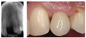
A 32-year-old male patient presented with a congenitally missing maxillary lateral incisor. Specialist orthodontic treatment had been completed elsewhere without any restorative interdisciplinary planning.

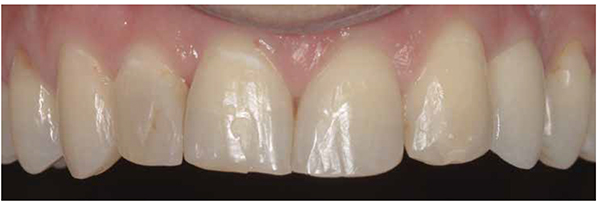
The UL3 was positioned in the UL2 space, and a pontic space was present in the UL3 area (Figure 1). A diminutive right lateral incisor was present. The overall dentition was minimally restored, and the pontic space mesiodistally was adequate for a canine width tooth (Figure 2). However, the interradicular space was limited and inadequate for replacement of this tooth with an average diameter implant, ideally suited for a canine tooth (Figure 3).

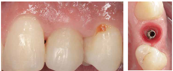
After a detailed discussion, the patient ruled out a resin-retained bridge and preferred to have the tooth replaced with a dental implant. Direct composite buildup was carried out to optimize the diminutive right lateral incisor and reshape the UL3 as the UL2.

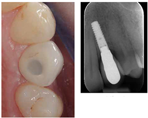
A narrow diameter, tissue-level type, Straumann® Roxolid® hydrophilic implant (3.3 mm diameter NNC) was placed in an ideal three-dimensional position for a screw-retained single-tooth restoration. Submerged healing was allowed for 6 weeks followed by implant exposure surgery.

Figure 5: Soft tissue sculpting is carried out over a few visits by modifying the provisional crown; Figure 6: The emergence of the restoration mimics a natural tooth, and the implant is ready
Figure 7: The shape of the impression coping does not correspond to the shape of the subgingival emergence. To facilitate the technician, impression coping should be customized; Figure 8: A hard-setting silicone bite registration material (Stone Bite®, Dreve Dentamid GmbH) is used directly in the mouth to capture the detail of the subgingival emergence
Figure 9: Customized impression coping is incorporated within the impression, and an ideal working cast can be created; Figure 10: Postoperative view. The final restoration, fulfilling all the pink and white esthetic criteria

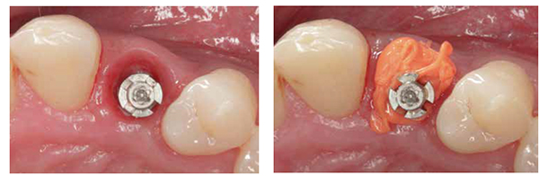
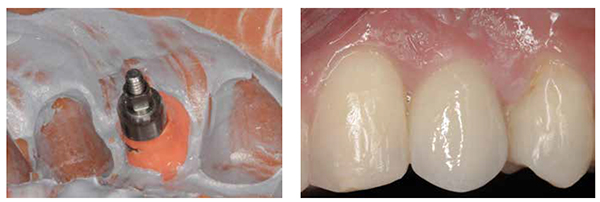
After a period of provisionalization to allow peri-implant soft tissue molding (Figures 4-6), impression coping was customized to facilitate the dental technician (Figures 7 and 8) to achieve an ideal emergence in the definitive restoration (Figure 9).

Conclusion
While the success of osseointegration is now an established fact, the challenge for clinicians is to deliver restorations that esthetically fulfill the objectives of treatment. This clinical case demonstrates how careful assessment and treatment planning can be predictably controlled to fulfill the treatment objectives.

A narrow diameter implant was used to replace a canine, which is now feasible to do predictably with the advent of stronger dental implant materials.

Despite the non-ideal orthodontic result, the final esthetic outcome with the dental implant and reshaping of teeth with directly bonded composite restorations yielded a great result for this patient (Figures 10 and 11).

Figure 11: Two-year follow-up. The imperceptible restoration despite malpositioned UL3 in the UL2 space
Figure 12: Definitive screw-retained restoration — the contours of which should snugly fit the subgingival emergence. The restoration should be neither over- nor under-contoured; Figure 13: Radiograph at 2 years, demonstrating well maintained bone levels

Author Info

Shakeel Shahdad, BDS, MMedSc FDS, RCSEd FDS (Rest Dent), RCSEd, DDS, is a consultant and honorary clinical senior lecturer in restorative dentistry at The Royal London Dental Hospital and Barts and The London School of Medicine and Dentistry, Queen Mary University of London. He is a specialist in restorative dentistry, periodontics, prosthodontics, and endodontics. He also works at Specialist Dental Services, a specialist referral practice at 7 Wimpole Street, London, England.

References

  1. Fürhauser R, Florescu D, Benesch T, Haas R, Mailath G, Watzek G. Evaluation of soft tissue around single-tooth implant crowns: the pink aesthetic score. Clin Oral Implants Res. 2005;16(6):639-644.
  2. Jemt T. Regeneration of gingival papillae after single-implant treatment. Int J Periodontics Restorative Dent. 1997;17(4):326-333.
  3. Tarnow DP, Cho SC, Wallace SS. The effect of inter-implant distance on the height of inter-implant bone crest. J Periodontol. 2000;71(4):546-549.

Stay Relevant with Implant Practice US

Join our email list for CE courses and webinars, articles and mores

Read our following terms and conditions before subscribing.

Terms and Conditions checkbox is required.
Something went wrong. Please check your entries and try again.

SUBSCRIBE TODAY

Implant Practice US is a leading dental journal and publication for dental implantology continuing education, oral implantology case studies, and more. Subscribe to Implant Practice US today!

ONLINE DENTAL CE

Earn dental continuing education credits as an Implant Practice US subscriber. Log in for online dental CE credits now!

Other Dental Publications

AGD PACE MedMark White

SUBSCRIBE TODAY

Implant Practice US is a leading dental journal and publication for dental implantology continuing education, oral implantology case studies, and more. Subscribe to Implant Practice US today!

ONLINE DENTAL CE

Earn dental continuing education credits as an Implant Practice US subscriber. Log in for online dental CE credits now!

Other Dental Publications

Copyright © 2024 Orthodontic Practice US - Dental Journal and Online Dental CE | MedMark LLC
15720 North Greenway Hayden Loop, Suite #9 Scottsdale, AZ 85260 | All rights Reserved | Privacy Policy | Terms & Conditions

Scroll to Top