Dr. Brad McAllister offers hope to a patient with a hopeless tooth
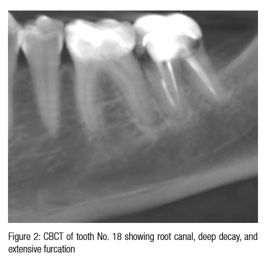
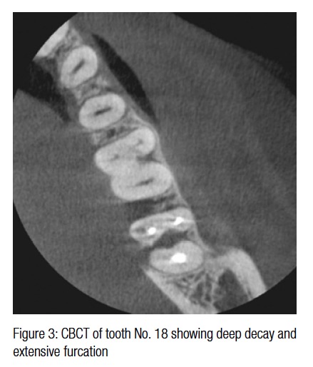
 A 50-year-old female presented with hopeless tooth No. 18 (Figure 1). Her medical and dental history were uneventful. This previously root canaled tooth was hopeless due to deep decay and extensive furcation involvement (Figures 2 and 3). In planning a size for placing an immediate Straumann® Roxolid® SLActive® Bone Level Tapered (BLT) Implant, a cross-sectional view was employed to measure the distance to the inferior alveolar nerve (Figure 4). Approximately 3 mm of bone was observed apical to the tooth root and superior to the nerve.
[userloggedin]
 The patient initiated a 1-week course of antibiotic therapy, which included 500 mg of amoxicillin 3 times per day, the day prior to her surgery. After local anesthetic was administered, a localized flap was elevated, and the tooth was sectioned and extracted taking care to preserve the buccal plate of bone.
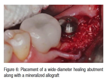
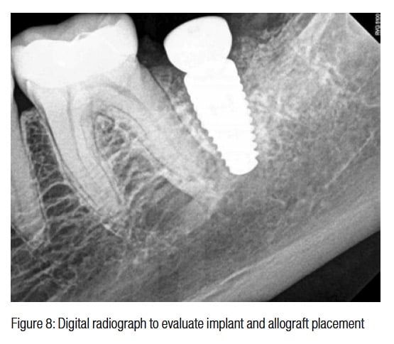
 The socket was degranulated, curetted, and rinsed with sterile saline prior to initiating any implant osteotomy preparation. The osteotomy was underprepared in diameter and the Straumann® Roxolid® SLActive® BLT implant (10 mm x 4.8 mm RC) was inserted with a torque of 35Ncm (Figure 5). The BLT implant design easily allowed for ideal placement and excellent initial stability. A wider diameter healing abutment was placed along with a mineralized allograft (Figure 6). The site was sutured to a tension-free primary closure with Vicryl® (Ethicon) 4-0 suture (Figure 7). A digital radiograph was taken to evaluate implant and bone graft placement (Figure 8).
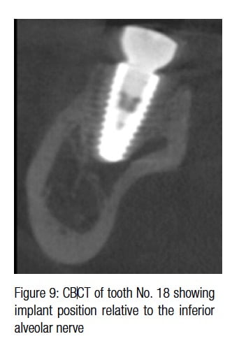
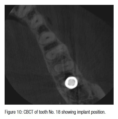
Since the inferior alveolar nerve positioning was not clear, a CBCT was taken to verify implant position relative to the inferior alveolar nerve (Figures 9 and 10). It was observed that nearly 2 mm of clearance was present. Initial healing was uneventful as shown in the 1-month healed clinical presentation (Figure 11).
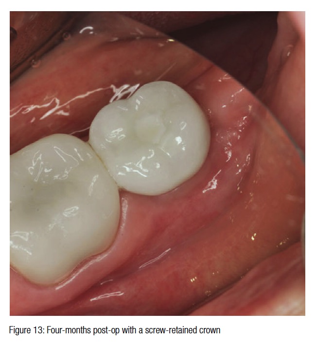
Following 4 months of healing, the final restoration of a screw-retained gold abutment with a crown consisting of porcelain fused to metal was fabricated and torqued to 35Ncm (Figures 12 and 13). The radiographic evaluation showed ideal restoration contours and excellent radiographic bone healing (Figure 14).
[/userloggedin]
[userloggedout][/userloggedout]
Stay Relevant With Implant Practice US
Join our email list for CE courses and webinars, articles and mores

