Dr. Ara Nazarian’s technique for immediate guided implant placement and provisionalization can help patients who require full-mouth extractions.
Dr. Ara Nazarian discusses his technique for questionable and/or non-restorable teeth requiring full mouth extractions
When a patient presents to your dental practice with questionable and/or non-restorable teeth requiring full mouth extractions, the biggest concern is whether or not implants can be placed at the same surgical visit, and if so, will the patient be able to walk out with fixed teeth. Using an implant, which allows you to load or progressively load so that this type of patient’s demands are met, allows you to position your practice to a whole new level. Of course, certain parameters must be met in order to facilitate this type of treatment. This includes, but is not limited to, the quality and quantity of bone, the presence of infection, the patient’s health, and the skills of the dental provider. Additionally, the selection of the most appropriate materials for the most ideal situation must be met.
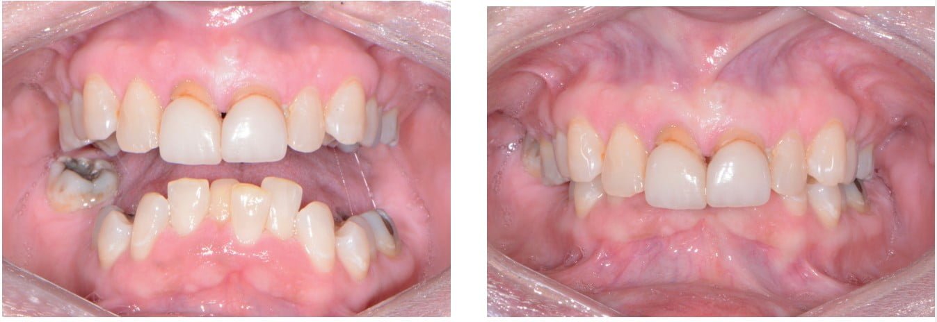
A patient presented to my practice for a consultation wanting to restore her dentition to proper form and function (Figure 1). She complained of generalized discomfort and mobility in these teeth apparently due to advanced periodontal disease. There were several teeth in both arches that had Class III mobility upon clinical examination. Also, there was hyper-eruption in the anterior mandibular dentition due to her jaw position with a deep impinging bite (Figure 2).
Planning
The clinical evaluation included information regarding lip length and support, existing tooth position of the natural teeth, occlusion, restorative space, and phonetics. In addition, digital images of frontal, side, and occlusal views of the dentition as well as facial shots were captured with a Nikon D7200 (PhotoMed).
A CBCT scan and panorex, using the CS 8100 3D (Carestream Dental) (Figure 3), were taken to accurately capture the information needed to properly treatment plan this case ensuring the most ideal outcome, especially since the patient had discussed how disappointed she was with her existing smile. Using the CS 3D imaging software, dental implants were virtually planned in key positions in both maxillary and mandibular arches (Figure 4).


To further develop a treatment plan, diagnostic model impressions were taken using Silginat® (Kettenbach) polyvinyl siloxane impression material, then poured up and forwarded to the dental lab. These models were then mounted on an articulator (Stratos 100, Ivoclar Vivadent) for further analysis in order to meet the patient’s esthetic and functional needs.
Financing options using a third-party payment option (LendingClub) were discussed with the patient. This discussion was a very important part of facilitating acceptance of her care, since it made the cost of treatment more feasible.
A 3D virtual treatment plan was further developed from our planning with the CS 3D imaging (Carestream Dental) software and integrating it with the photos and models. A virtual online integrative meeting with the dental lab allowed for a comprehensive review of the assembled digital and clinical information formulating an optimal treatment plan that would fulfill the necessary requirements for esthetics, form, and function. Within a short amount of time, the dental lab had fabricated all the necessary guides for positioning, leveling, drilling, and implant placement in addition to the PMMA provisional restorations and backup dentures (Figure 5).

It is my belief that surgical guides in implant dentistry increase the predictability of treatment outcomes as well as making the clinician extremely efficient. In the past, implant placement routinely occurred by freehand, but this technique heightened the risk of damage to anatomic structures while lengthening the duration of the surgery. I personally feel surgical guides give clinicians more confidence to accurately place implants in every case whether you are a general practitioner or specialist. Precision surgery reduces stress, decreases liability, and leads to a better outcome for the patient.
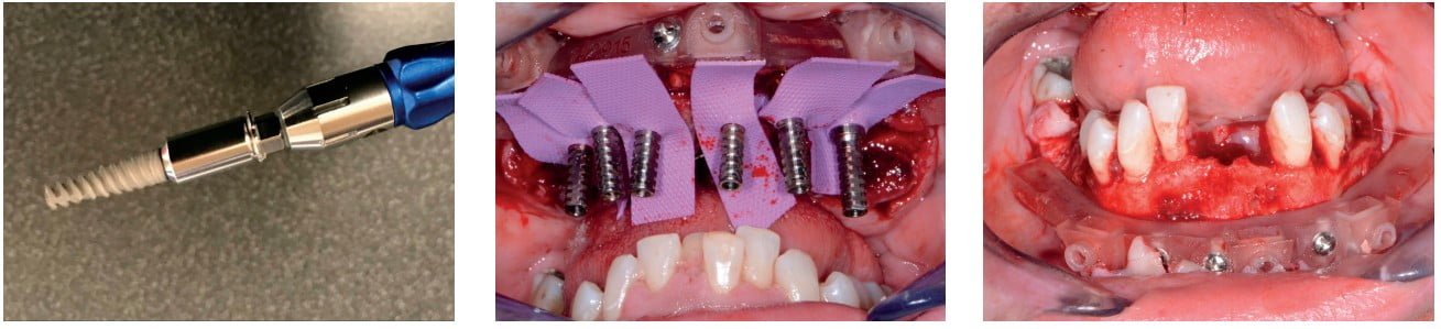
The implants that would be utilized for this case were the Adin Touareg™ OS dental implants (Figure 6). The Touareg™ S & OS Spiral Implants are tapered with a spiral tap that condenses the bone during placement for immediate stability. There are two large variable threads and a tapered design for accurate implant placement, self-drilling, improved esthetics, and better load distribution. They feature a special round-shaped apex that pushes the bone graft with minimal harm to anatomic structures. In addition, Touareg™ OS implants feature Adin’s biocompatible and osseoconductive OsseoFix™ implant surface. This has proven to achieve the desired roughness levels for optimal osseointegration, attains the highest implant surface purity levels, and increases the success rate of bone-to-implant contact (BIC).
Once the virtual plan was orchestrated and fully confirmed, the next appointment would be the planned surgery with all the necessary components for the guided surgical approach. The patient was appropriately sedated with IV medications, and local anesthesia was administered in both arches. The tissue was then reflected using the Reflector (GoldenDent™) instrument so that the bone-leveling surgical guide would be fully seated and fixed with its respectful retention screws (Figure 7). Following the positioning of the surgical guide, the maxillary teeth were atraumatically extracted utilizing the Physics Forceps® (GoldenDent). Once the appropriate bone leveling was accomplished with the surgical handpiece, the implant surgical guide (Figure 8) was positioned into the bone-leveling guide, and the sites for the implants was initiated with a designated pilot drill in the Adin Guided Surgery Drill Kit (Figure 9).

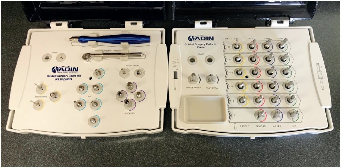
Using precise orientation, depth, and direction, Adin’s Guided Surgical Kit provides fast, effective, and predictable preparation and placement of dental implants for dental practioners. It also features easy-to-follow layouts containing self-centering drills with built-in stoppers. The unique design of the Adin Guide ActiveFlow™ Irrigation Technology directs cooling saline through the guide, ensuring that irrigation reaches the bone, reducing the possibility for bone-heating throughout the procedure.
Sequential drill preparation was initiated utilizing the Mont Blanc® surgical handpiece and the Aseptico surgical motor (AEU 7000) at a speed of 800 rpm with copious amounts of sterile saline. Once the osteotomies were complete, the drivers in the Adin Guided Kit (Figure 10) were used to place the dental implants with precise timing so that the flat portion of the internal hex was positioned ideally for the receiving multiunit abutments.
A baseline ISQ reading was taken of these implants utilizing the Penguin (Aseptico) RFA unit. Since the initial readings were all above 70 and the quality of bone after leveling was good, multiunit abutments (Adin) were tightened into the Touareg OS (Adin) dental implants at 30, followed by temporary cylinders at 15 Ncm.
Any residual areas around the implants or in the sockets were grafted with a cortical mineralized and demineralized bone-grafting material (GoldenDent) to optimize the area for regeneration.
The prefabricated immediate provisional arch restorations with predrilled access openings were inspected before being trying-in.
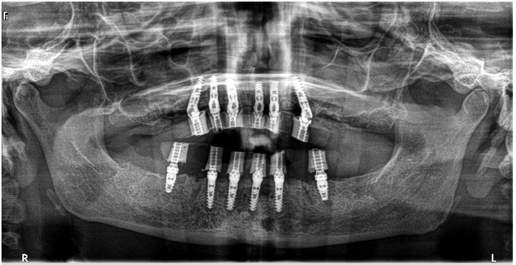
The maxillary provisional restoration was tried-in to verify a passive fit over the temporary abutments. Once confirmed, trimmed rubber dam pieces were placed to avoid the restoration (Figure 11) from locking on during the relining procedure with Rebase III Fast Set (Tokuyama®) hard reline material. After the material polymerized, the immediate provisional restoration was removed, and any access material removed with the Torque Plus (Aseptico) lab handpiece and acrylic bur (Komet). The same procedures were accomplished in the lower arch (Figures 12-14). Once trimmed and polished, the provisional arch restorations were seated and tightened with a torque wrench at 15 Ncm (Figure 15). The access openings were filled ¾ of the way with Teflon® tape followed by Cavit™ (3M™) filling material. A postoperative panorex radiograph was taken immediately after the surgery (Figure 16).

A few days later, the patient returned for her postoperative appointment with very little discomfort, swelling, or bruising. She was very pleased with her new upper- and lower-fixed provisional restorations. The occlusion was further checked and adjusted to confirm there were no interferences in lateral or protrusive movements. The next step in her treatment would consist of full arch impressions for the definitive restorations approximately 4 to 5 months postoperatively.
Conclusion
Having the ability to take a patient from start to finish in a fewer amount of appointments within your practice allows you to position yourself as a provider who can fulfill your patients’ surgical and restorative needs. With the proper training and appropriate materials, a dental provider may provide extraction, grafting, and implant placement within one appointment at one location. This type of service not only allows you to reduce the amount of visits for the patients, but also helps maintain the cost to the patients, since they are not seeing multiple dental providers. Most importantly, this enables the dental provider full control of the surgical and prosthetic outcome. Depending on the patients’ desires, the clinical conditions of the oral environment present, and the skills of the provider, a dentist may choose to extract teeth, level bone, and graft with guided dental implant placement within his/her dental practice.
Dr. Nazarian also uses a guided implant placement technique in his article “Full arch fixed provisionals with dual stabilization dental implants.” Read about how his patient received treatment with fewer appointments: https://implantpracticeus.com/full-arch-fixed-provisionals-dual-stabilization-dental-implants/
 Ara Nazarian, DDS, DICOI, maintains a private practice in Troy, Michigan, with an emphasis on comprehensive and restorative care. He is a Diplomate in the International Congress of Oral Implantologists (ICOI). Dr. Nazarian has conducted lectures and hands-on workshops on esthetic materials, grafting, and dental implants throughout the United States, Europe, New Zealand, and Australia.
Disclosure: Dr. Nazarian is the creator of the Reflector instrument. He has reported no other compensation for other products mentioned in this article.
Stay Relevant With Implant Practice US
Join our email list for CE courses and webinars, articles and mores

