Dr. Salah Huwais discusses how osseodensification facilitates ridge expansion with enhanced implant stability
Introduction
The medical profession has, with certain exceptions, adapted commercially available instruments that have been developed for drilling other materials (Jackson, et al., 1989). For more than a decade, clinicians have been asking for improvement in bone drilling and preparation (Natali, et al., 1996).
Standard drill designs used in dental implantology are made to excavate bone to create room for implant placement. They cut away bone effectively but typically do not produce a precise circumferential osteotomy. Osteotomies may become elongated and elliptical due to the chatter of the drills.
[userloggedin]
In these circumstances, the implant insertion torque is reduced leading to poor primary stability and potential lack of integration. Furthermore, osteotomies drilled into narrow bone locations may produce dehiscence, buccally or lingually, which also reduces primary stability and will require an additional bone grafting procedure adding cost and healing time to treatment.
When standard drills extract enough bone to let strains in the remaining bone to reach or exceed the bone micro-damage (MDX) threshold, the bone-remodeling unit (BMU) needs more than 3 months to repair the damaged area, so maintaining bone bulk will enhance healing and shorten the healing period (Frost, et al., 1998).
Unlike traditional bone drilling technologies, osseodensification does not excavate bone tissue. Rather, it preserves bone bulk, so bone tissue is simultaneously compacted and autografted in an outwardly expanding direction to form the osteotomy. It is accomplished by using proprietary densifying burs. When the densifying bur is rotated at high speed in a reversed, non-cutting direction with steady external irrigation (Densifying Mode), a dense compacted layer of bone tissue is formed along the walls and base of the osteotomy (Meyer, Huwais, et al., 2014).
The goal in implant placement is to achieve primary implant stability. It is well established that implant stability is critical for osseointegration (Albrektsson, et al., 1986, Meredith, et al., 1998). This is more important in recent days due to popular immediate/early loading protocols being implemented into treatment by many clinicians. Removing bone bulk is contrary to achieving the primary stability desired.
Implant primary mechanical stability is directly related to surrounding bone quality and quantity. Maintaining and preserving bone during osteotomy preparation leads to increased primary mechanical stability, increased bone to implant contact (BIC), which then enhances implant secondary stability, and accelerates healing (Seeman, et al., 2008, Todisco, et al., 2005, Trisi, et al., 2009).
Case report
Osseodensification facilitates mandibular ridge expansion and placement of two implants.
The patient is a 62-year-old male presented with missing teeth Nos. 19, 20, and 21. Clinical and radiographic examination revealed a significant alveolar ridge resorption, which resulted in a Seibert Class I, ridge deficiency (Figure 1). The patient’s medical history was noncontributory. Treatment options with their potential risks and benefits were presented to the patient. A final treatment plan was finalized to utilize placement of two implants to receive two abutments for a fixed prosthesis to restore teeth Nos. 19, 20, and 21. Consent was given by the patient to utilize osseodensification site preparation for ridge expansion with immediate implant placement and possible additional buccal bone grafting if needed.

 The lower left area was anesthetized using infiltration method with 1.8 ml 4% Septocaine® (Septodont) with 1:100,000 epinephrine. Once anesthetized, a crestal incision was done, and a full thickness flap was reflected to reveal 2.5 mm-3.0 mm crestal alveolar ridge width, which was confirmed by direct measurement (Figure 2).


 The site preparation for two implants in the areas of Nos. 19 and 21 began with site marking. Then, a 1.5-mm initial pilot osteotomy was created with a pilot drill rotated at 1200 RPM in a clockwise rotation (CW) to a depth of 13 mm utilizing a high-speed surgical handpiece and a surgical motor (W&H) (Figure 3). Using paralleling pins, an X-ray was taken to confirm the angulation between the adjacent teeth and the implants.
Once implant positions were confirmed, a horizontal ridge split to a 10-mm depth was created using Piezosurgery® (Piezosurgery Incorporated) to allow further buccal plate flexibility. Osseodensification with ridge expansion started with Densah™ Bur VT1525 (Versah™, LLC) rotating in a non-cutting counterclockwise (CCW) direction at 1200 RPM (Densifying Mode) to expand the osteotomy to 2.5 mm, utilizing a high-speed surgical handpiece and a surgical motor (W&H) (Figure 4).
Then Densah™ Bur VT2535 (Versah, LLC) running in a non-cutting counterclockwise (CCW) direction at 1200 RPM (Densifying Mode), utilizing a high-speed surgical handpiece and a surgical motor (W&H), was used to expand osteotomies in the area of implant Nos. 19 and 21 (Figure 5). Mandibular osteotomies were expanded to 3.5 mm without any bone dehiscence, which then allowed for total implant length placement in autogenous bone without any thread exposure (Figure 6).
Two 3.7/13 Tapered Screw-Vent implants (Zimmer®) were placed with an insertion torque of 40-50 Ncm. Both implants total lengths were covered with autogenous bone. Less than 1.0 mm crestal-buccal bone thickness in area of implant No. 21 was noted (Figure 7). Implant stability was measured with an (Osstell®) ISQ implant stability meter, which uses resonance frequency analysis. In this particular case, buccal-lingual ISQ readings in the areas of Nos. 19 and 20 were 78 and 49, respectively. Several studies have been conducted on resonance frequency analysis (RFA) measurements and the ISQ. They provided valid indication that accepted stability range is above ISQ 50 and recommended loading at ISQ 67-68.
Due to an ISQ reading of 49 in the mesial implant No. 21 and less than 1.0 mm of crestal-buccal bone thickness remaining after osseodensification, the decision was made to augment the buccal plate with a bone graft (Figure 8).
Healing cover screws were placed and Puros Demineralized Bone Matrix (Zimmer®) was used as allograft to augment the mandibular buccal bone plate post implant placement. Full flap closure with mattress suture was achieved (Figures 9 and 10).
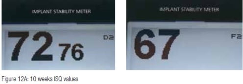
Eight weeks post placement, implants were uncovered through shallow crestal incision. Healing abutments were placed. Buccal-lingual ISQ readings obtained at week 10 were 76/72, 67 for implant Nos. 19 and 20, respectively. The implants’ high insertion torque with maintained gain in ISQ had allowed us to consider an early restorative phase initiation.Thus, at 10 weeks, when the ISQ reading reached ≥ 67, the patient was referred back to his restorative dentist for the restorative phase.Fourteen weeks post implant surgery, a fixed prosthesis retained by implants Nos. 19 and 21 was delivered.
Supportive and follow-up care
The patient returned in 1 year for clinical and radiographic follow-up. Examination revealed healthy hard and soft tissue with no sign of inflammation or infection. Radiographic examination revealed maintained crestal bone level and bone density. Clinical examination revealed slight soft tissue recession in the area of implant No. 21. This soft tissue height reduction is common post GBR or ridge augmentation procedures. Four months’ interval supportive periodontal treatment was initiated with yearly radiographic examination for implant Nos. 19 and 21.


 In this case, osseodensification utilizing the Densah™ Bur technology had facilitated ridge expansion with maintained alveolar ridge integrity, allowing for total implant length placement in autogenous bone with adequate primary stability. Despite compromised bone anatomy, osseodensification preserved bone bulk and promoted a shorter waiting period to the restoration.
Ordinarilly, a case similar to this patient would progress through three phases of treatment over 30-50 weeks:
- Ridge augmentation phase (6-9 months) to increase ridge width with either block grafting or guided bone regeneration
- Implant placement and healing phase (2-3 months)
- Restorative phase
The question remains, why do we build bone bulk to then extract it later and wait months for implants to heal? It is time to think about bone preservation to enhance its ability to heal faster, regardless of implant macro- or micro-geometry.
Conclusion
Osseodensification is a novel, bio-mechanical, non-excavation osteotomy preparation method. Unlike traditional drilling, osseodensification utilizes proprietary high-speed densifying burs to compact and autograft bone in its plastic deformation phase. The result is an expanded osteotomy with preserved and condensed bone tissue that maintains alveolar ridge integrity and allows for implant placement with enhanced stability.
References
Jackson CJ, Ghosh SK. On the evolution of drill-bit shapes. Journal of Mechanical Working Technology. 1989;18(2):231-267.
Natali C, Ingle P, Dowell J. Orthopaedic bone drills-can they be improved? Temperature changes near the drilling face. J Bone Joint Surg Br. 1996;78(3):357-362.
Frost HM. A brief review for orthopedic surgeons: fatigue damage (microdamage) in bone (its determinants and clinical implications). J Orthop Sci, 1998;3(5):272-281.
EG, Huwais S. Osseodensification Is A Novel Implant Preparation Technique That Increases Implant Primary Stability By Compaction and Auto-Grafting Bone. American Academy of Periodontology. [abstract]. San Francisco, CA. 2014.
Albrektsson T, Zarb G, Worthington P, Eriksson AR. The long-term efficacy of currently used dental implants: A review and proposed criteria of success. Int J Oral Maxillofac Implants. 1986;1(1):11-25.
Meredith N. Assessment of implant stability as a prognostic determinant. Int J Prosthodont. 1998;11(5):491-501.
Todisco, M, Trisi P. Bone mineral density and bone histomorphometry are statistically related. Int J Oral Maxillofac Implants. 2005. 20(6):898-904.
Seeman E. Bone quality: the material and structural basis of bone strength. J Bone Miner Metab. 2008;26(1):1-8.
Trisi P, Perfetti G, Baldoni E, Berardi D, Colagiovanni M, Scogna G. Implant micromotion is related to peak insertion torque and bone density. Clin Oral Implants Res. 2009;20(5):467-471.
[/userloggedin]
[userloggedout][/userloggedout]
Stay Relevant With Implant Practice US
Join our email list for CE courses and webinars, articles and mores
