Educational aims and objectives
This clinical article aims to present a case study where the patient’s desire to avoid wearing a temporary denture formed a fundamental part of the treatment-planning process.
Expected outcomes
Implant Practice US subscribers can answer the CE questions by taking the quiz to earn 2 hours of CE from reading this article.
Take the quiz by clicking here.
Correctly answering the questions will demonstrate the reader can:
- See diagnosis and treatment of a patient who was reluctant to wear dentures that resulted ineffective but flexible treatment planning.
- Note how the patient’s esthetic analysis affected treatment.
- Identify some special tests that were conducted to help deduce the diagnoses.
- Realize the role of CBCT imaging in diagnosis and treatment planning.
- Recognize several post-extraction treatment options for this patient.
Immediate surgery and implant-supported prosthetics were pursued when the patient discussed her wish to avoid a traditional denture.
Dr. Dipesh Parmar details a case where the patient’s need to avoid a denture shaped the progression of treatment
In pursuit of esthetic and functional perfection, clinicians often fail to see beyond these goals and fully appreciate the psychological impact that the removal of teeth has on our patients. The following case demonstrates how dental implants can be used to restore edentulous arches. The case is about not only the rehabilitation of a patient’s dentition, but also the management of her reluctance to lose her remaining teeth, the building of rapport and trust, the management of expectations, and ultimately, the restoration of a person’s confidence and ability to socially re-engage.
The implant surgery and immediate prosthetic work was carried out by Dr. Manoj Parmar. The work related to the definitive prostheses was undertaken by Dr. Dipesh Parmar.
History and examination
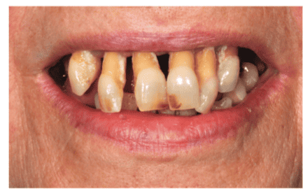
A 59-year-old retired teacher attended the practice, expressing her unhappiness with the appearance of her teeth and the psychosocial effect it was having on her. Eating was becoming increasingly difficult due to the lack of opposing teeth and the advanced mobility of the remaining units. Due to embarrassment, she had not visited a dentist for more than 5 years; however, the extreme lack of self-confidence eventually forced her to get her concerns addressed. She complained of no pain or discomfort from her teeth.
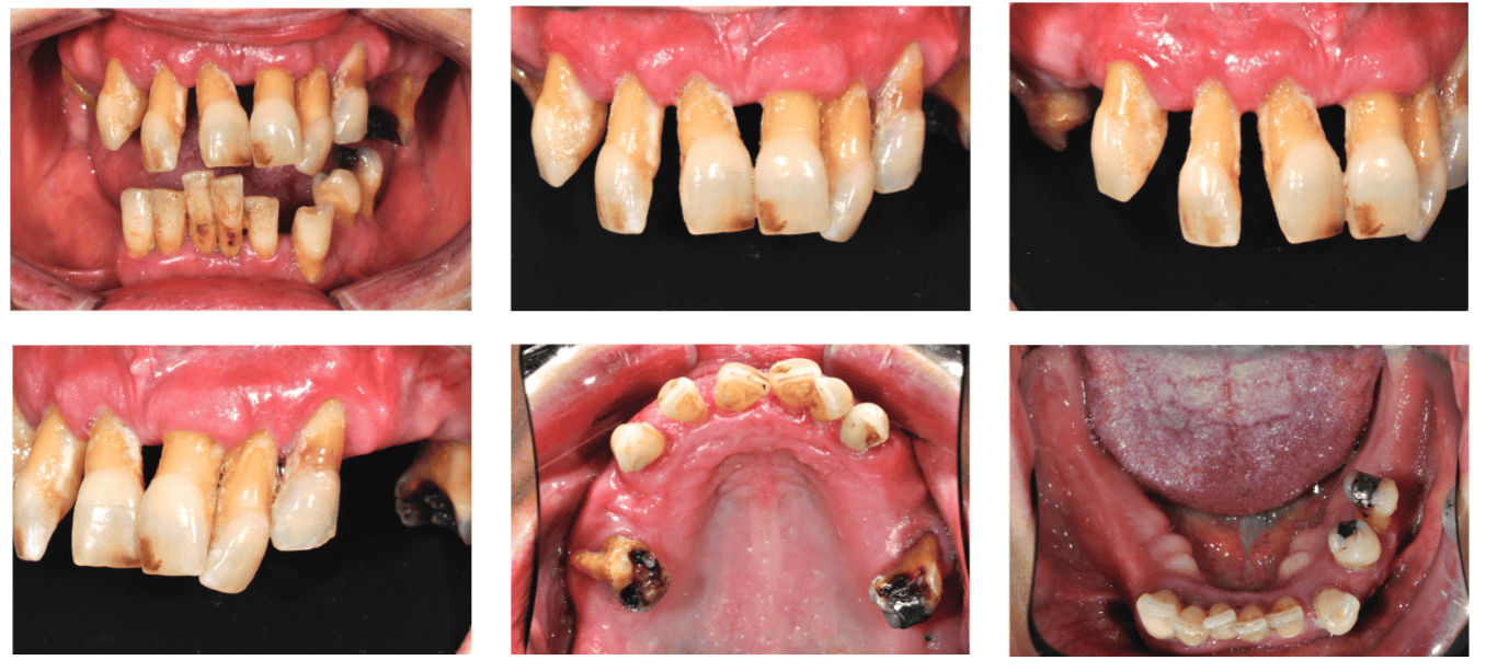
Medically, she suffered from mild asthma that was controlled with salbutamol. No allergies were recorded. She was a nonsmoker, and her alcohol intake was minimal. Family history revealed early loss of teeth of teeth on her maternal side of the family. A diet history highlighted frequent sugar intake. Extraoral examination revealed nothing abnormal. Intraorally, the soft tissues were healthy. Her oral hygiene was graded as poor with evidence of generalized biofilm deposits and associated gingival inflammation and suppuration. She presented with medium-to-thick gingival biotype, and the BPE was recorded as 444/-44.
Occlusal analysis proved to be difficult due to numerous missing units and the effects of the periodontitis on the remaining dentition.
However, she presented with a Class II Division I incisor relationship on a Class I skeletal base. Due to the lack of teeth, lateral excursive movements had no alternative but to be canine-guided. Centric occlusion was not coincident with centric relation with the first contact between teeth 26 and 35.
Most likely attributed to the lack of posterior support, she presented with an increased overjet and an increased and complete overbite. She had no history of parafunction; however, mobility and fremitus were present on all teeth.
Esthetic analysis highlighted the following:
- Midlines — unacceptable. Dental midline shifted to left
- Lip line — high
- Smile line — discontinuous
- Incisal edge position — central incisors and canines within normal limits. Lateral incisors unacceptable
- Buccal corridor —unacceptable
- Central incisor dimensions — acceptable (clinical crown only, roots excluded)
- Tooth-to-tooth proportions — acceptable
- Axial inclinations — unacceptable
- Gingival esthetics — unacceptable due to effects of periodontitis
- Connector heights — unacceptable
- Embrasure form — unacceptable
- Labial anatomy — within normal limits
- Color — unacceptable
Special tests
The following special tests were conducted to help deduce diagnoses:
- Full-mouth detailed pocket charting was carried out, with bleeding indices, plaque indices, and mobility grading. All teeth scored either Grade 2 or 3 mobility.
- Thermal and cold pulp tests were carried out. Teeth UR6, UL3, UL6, LL4, and LL5 tested negatively.
- Percussion tests were also applied. Teeth UR6, UL3, UL6, LL4, and LL5 tested tender to percussion in the apical direction. All other teeth responded positive to lateral percussion tests.
- Periapical radiographs were taken of the lower teeth to assess bone levels and apical pathology. Radiographic assessment highlighted 80%-100% bone loss on all mandibular teeth with evidence of vertical and crescentic bone loss patterns. Apical pathology was noted on UR6, UL3, UL6, LL1, LL4, LL5, and LR1.
- A maxillary CBCT scan was taken by the implant surgeon to allow assessment for implant treatment.
- The full series of British Academy of Cosmetic Dentistry (BACD)-recommended clinical photographs were taken. Figures 1-7 show a selection of these.
Diagnoses
Following thorough clinical and radiographic assessment, coupled with multiple special investigations, the following diagnoses were made:
- Advanced generalized chronic perio-dontitis affecting all units
- UR6 and UL6 — Asymptomatic perio-endo lesions with evidence of gross caries
- LL4 and LL5 — Asymptomatic perio-endo lesions
- UL3 — Lost restoration and distal caries
- UL7 — retained root remnant
- Multiple missing units with resultant loss of function
- Compromised esthetics
Treatment objectives and options
The main objective of dental treatment for this patient was to re-establish form, function, and esthetics of her masticatory system. In addition, it was imperative to motivate the patient to improve her oral hygiene and rebuild her self-confidence and faith in dentistry. It was evident that the remaining teeth had very poor prognosis, and the most appropriate treatment plan to fulfil the objectives was full-mouth clearance followed by fixed or removable prostheses.
The following post-extraction treatment options were discussed with the patient.
Removable prostheses
- Acrylic (with or without cobalt chrome) tissue-borne prostheses
- Implant-supported overdentures
Fixed prosthesis
- Hybrid bridge on four or more implants (immediate or delayed loading)
- Ceramic-metal bridge(s) on six or more implants
The patient’s initial wishes were to have a fixed maxillary implant-retained ceramic-metal bridge and a conventional mandibular acrylic prosthesis. She wanted to achieve the best esthetic outcome in the upper arch and, due to the financial implications of this, was happy to accept a compromise solution for the lower arch.
Further discussions took place regarding the use of dissimilar materials in opposing arches and that the full benefits of having a fixed solution in the upper arch could not be attained while having a conventional prosthesis in the lower arch.
Planning
Following detailed discussion, enhanced by the use of previous case studies, it was decided with the patient that the most appropriate plan would be extraction of the remaining teeth with provision of immediate replacement dentures (IRDs). This would be followed by a maxillary implant-retained hybrid bridge on four implants and a man-dibular implant-supported overdenture on two implants.
Treatment
Phase 1: Lower-arch clearance
For the purposes of mental conditioning, it was planned to treat each arch separately starting with the lower at the
patient’s request.
Measurements were taken for the construction of a lower complete denture, which was fitted at the final visit immediately after the removal of all mandibular teeth. The granulation tissue from all the sockets was also curetted to facilitate optimal healing.
At the follow-up appointments, the patient expressed her discontent with the poor retention and stability of the lower prosthesis, despite its relining with tissue conditioners. This reinforced not only the decision to have an implant-supported denture for the lower arch, but also her desire not to go through same process for the upper arch.


Phase 2: Upper-arch planning and treatment
In order to respect the patient’s decision not to have a clearance and an IRD, the treatment plan had to be revised. Although still in agreement for the definitive prosthesis to be a hybrid bridge, the only way to avoid an IRD was to extract the teeth, place the implants, and immediately load with a temporary bridge all on the same day.
To enable us to do this, the following plan was created:
- Patient to undergo hygiene therapy to remove all plaque deposits, arrest active disease, and reduce perio-dontal inflammation.
- Take a CBCT scan of the upper arch with a radiographic stent to assess bone levels for implant planning.
- Remove any teeth that were infected or unsavable.
- Construct an upper full denture that would be converted into a temporary implant bridge or used as an IRD if immediate loading was not possible (Figure 8).
- Create a copy of the denture in clear acrylic to be used as a surgical guide.
The CBCT revealed that there was sufficient bone for the placement of four implants. Arrangements were then made for the extraction of the UL3 due to advanced apical pathology, followed by impressions to begin the construction of the upper complete IRD. To reduce the risk of accidental extraction of the incisors while taking impressions, the teeth were splinted using flowable composite, and the embrasures were occluded using temporary inlay material, which was subsequently easily removed.
The periodontal treatment reduced the inflammation of the gingivae. Once the dentures were fabricated, the next phase was surgery.
The complete denture was made to full sulcus depth and full palatal coverage to allow seating once the teeth were removed. However, the flanges were hollowed-out apical to the teeth to facilitate easier removal of the flange when converting the denture to a bridge.
The surgical stent mirrored the design of the denture with the addition of a channel cutout along the occlusal surfaces of the teeth to set the prosthetic profile within which the implants would be placed.
It was stressed to the patient that in the event that primary stability was not achieved, she would need to wear the full denture until osseointegration was optimal.
Following local anesthesia, the remaining upper teeth were separated and extracted. The denture was tried in and checked for fit, following which occlusion was checked against the lower denture and adjusted accordingly. The transition zone between the prosthesis and the gingivae was checked, albeit with difficulty due to the anesthesia.
A crestal incision was made approximately from UR6 to UL6 with small relieving incisions at either end. A full thickness flap was raised to expose the ridge. Sufficient ridge width was present as predicted from the CBCT. A large round bur was used to flatten the ridge, which also would move the transition line further apically, allowing it to be better hidden by the upper lip. The extraction sockets were thoroughly curetted.
A pilot osteotomy was made in the midline to facilitate the positioning of the Malo implant guide that facilitates the placement of the implant at the correct angulation. The surgical guide was repeatedly located to ensure that implants were placed within the prosthetic envelope.
The implants were placed using a standard surgical technique following the Malo protocol (Figure 9). Peri-treatment periapical radiographs were taken to ensure the anterior wall of the maxillary sinus was not violated. Once the implants were placed, angled abutments were placed on top and adjusted to make all four emergence profiles as parallel as possible while still being within the prosthetic envelope.
Once positions were confirmed, the abutments were torqued to 35 Ncm as recommended by the implant system. Two 30° angled abutments were used for the posterior implants, and two 15° angled abutments were used for the anterior implants.
The denture was lined with red beading wax on the fit surface. It was then transferred in to the mouth and seated until the abutments indented the wax indicating the position of the implants (Figure 10). Holes were then cut in the denture in these positions. Temporary metal cylinders were attached to the abutments and the fit of the denture checked over them. Biomaterials were used to fill any remaining sockets and voids in the bone with subsequent closure of the flap with 4.0 Vicryl® (Ethicon®) sutures (Figure 11).
Cold-cured chairside acrylic was used to tack the denture onto the temporary cylinders. The cylinders were then unscrewed from the abutments and removed with the denture, which was sent to the lab with the lower denture and bite registration for finishing and conversion into a temporary bridge. Healing caps were placed over the abutments while the bridge was being prepared at the laboratory.
When the bridge was returned, the healing caps were removed and the temporary bridge was seated and torqued down to finger pressure. Access holes were occluded with PFTE tape and temporary inlay material. The final occlusion was checked and adjustments made to ensure evenly distributed contacts conforming to the appropriate occlusal scheme.
The patient was reviewed a week later with evidence of healing. The bridge was removed along with remaining sutures. The prosthesis was cleaned, and the patient was provided with a water flosser to help with hygiene.
Phase 3: Lower-arch implant placement
Approximately a month after the upper-arch surgery, the patient was invited back to have two implants placed in the lower arch. Following local anesthesia, two individual flaps were raised to allow implant placement.
Holes were cut in the denture, which in turn was used as a surgical guide to help make the osteotomies in the correct positions and angulation. Cover screws were placed and the flap closed to allow submerged healing. The patient continued to wear the lower denture during the healing period.
Phase 4: Final prosthesis
A period of 3 months was allowed for complete integration of the implants before continuing with the prosthetic phase of the treatment. The lower implants were exposed and the cover screws replaced with healing abutments. The upper bridge was removed (Figure 12), followed by upper and lower primary impressions for fabrication of custom acrylic trays. The bridge was then replaced following retorquing of the abutments.
A master abutment-level open tray impression was taken of the upper arch using monophase and light-bodied PVS impression materials (Figure 13). A lower implant-level master impression was taken in heavy-bodied and light-bodied PVS impression materials. The increased viscosity of the silicone putty allows for a muco-displacing impression to be taken, which in turn would result in a snug fit of the overdenture to the ridge.


Wax occlusal registration rims were used to record centric relation at the desired vertical dimension as per conventional full denture construction, followed by a facebow transfer. A verification jig was used to confirm the accuracy of the upper master impression (Figures 14-16). The verification jig did not seat fully; thus, the rim was sectioned close to the implant where the discrepancy was, which allowed passive seating of the two parts. These were then rejoined using acrylic resin and returned to the lab for repositioning of the analogue on the master cast.
A cast metal framework was constructed (Figure 17) and tried intraorally with a passive fit being confirmed (Figure 18) as it was on the model. A try-in of both upper and lower prostheses was undertaken, and the teeth proved to be too short. The patient also requested more irregularity in the positioning of the teeth.
A second try-in with longer teeth was conducted (Figure 19); however, on this occasion the try-in was built onto the metal framework to allow accurate assessment of the occlusion, esthetics, and phonetics.
The position of the transitional line was also evaluated and confirmed not to be visible when the patient force-smiles. The patient consented to progress to the definitive restorations (Figures 21-22).
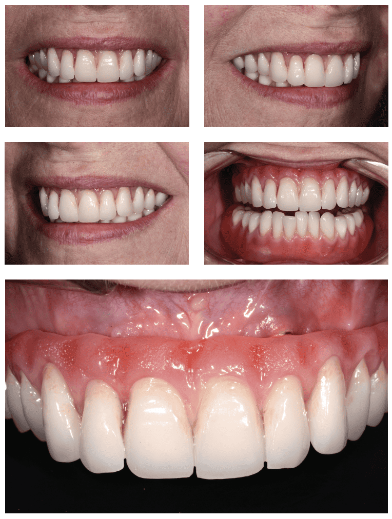
The temporary prostheses were removed, and the final bridge was fitted with access holes sealed with polytetrafluoroethylene (PTFE) tape and composite resin. Kerator® abutments were placed onto the lower implants, and the matrices were cold-cured into the lower denture. Written and verbal implant maintenance advice was provided to the patient. Figures 23 to 27 show the final result.
Conclusion
Full-mouth clearance cases can be extremely challenging, not only in terms of the surgical and restorative skills required to rehabilitate the patient, but also the management of patient expectations to deliver a result that exceeds the patient’s expectations as opposed to falling short of them. This was a truly rewarding case for the clinician but a life-changing one for the patient.
If a patient expresses the intention to avoid a traditional denture, sometimes a hybrid solution will fulfill his/her needs. Read Dr. Charlotte Stilwell’s CE article, titled, “Removable partial dentures and strategic implant placement.”
References
- Thomason JM, Feine J, Exley C, et al. Mandibular two implant-supported overdentures as the first choice standard of care for edentulous patients — the York Consensus Statement. Br Dent J. 2009;207(4):185-186.
- Chee W, Jivraj S. Treatment planning of the edentulous mandible. Br Dent J. 2006;201(6).:337-347.
- Chiapasco M. Early and immediate restoration and loading of implants in completely edentulous patients. Int J Oral Maxillofac Implants. 2004;19(suppl):76-91.
- Crespi R, Vinci R, Capparé P, Romanos GE, Gherlone E. A clinical study of edentulous patients rehabilitated according to the “all on four” immediate function protocol. Int J Oral Maxillofac Implants. 2012;27(2):428-434.
- Del Fabbio M, Bellini CM, Romeo D, Francetti L. Tilted implants for the rehabilitation of edentulous jaws: a systematic review. Clin Implant Dent Relat Res. 2012;14(4):612-621.
- Gallucci GO, Benic GI, Eckert SE, et al. Consensus statements and clinical recommendations for implant loading protocols. Int J Oral Maxillofac Implants. 2014; 29(suppl):287-290.
- Jivraj S, Chee W. Transitioning patients from teeth to implants. Br Dent J. 2006;201(11):699-708.
- Jivraj S, Chee W, Corrado P. Treatment planning of the edentulous maxilla. Br Dent J. 2006;201(5):261-279.
- Malo P, Rangert B, Nobre M. All-on-4 immediate function concept with Brånemark System implants for completely edentulous maxillae: a 1-year retrospective clinical study. Clin Implant Dent Relat Res. 2005;7(suppl):S88-S94.
- Malo P, de Araújo Nobre M, Lopes A, Francischone C, Rigolizzo M. “All-on-4” immediate-function concept for the completely edentulous maxillae; a clinical report on the medium (3 years) and long-term (5 years) outcomes. Clin Implant Dent Relat Res. 2012;14(suppl 1):e239-e150.
- Schimmel M, Srinivasan M, Herrmann FR, Müller F. Loading protocols for implant-supported overdentures in the edentulous jaw: a systematic review and meta-analysis. Int J Oral Maxillofac Implants. 2014;29(suppl):271-286.
- Stoker GT, Wismeijer D. Immediate loading of two implants with a mandibular implant-retained overdenture: a new treatment protocol. Clin Implant Dent Relat Res. 2011;13(4):255-261.

 Dipesh Parmar, BDS, graduated from the University of Birmingham (UK), obtaining numerous prizes and practices in Birmingham and focusing on minimally invasive esthetic reconstructions and orthodontic treatment. He has held a clinical lecturer teaching post at Birmingham Dental School and has won multiple awards at the UK Aesthetic Dentistry Awards, including Best Aesthetic Dentist UK 2018. He lectures at an international level on direct resin restorations. He has been awarded Diplomate status in orthodontic dentistry from the University of Warwick.

