Drs. Orcan Yüksel, Bernhard Giesenhagen, and Kris Chmielewski demonstrate a new method for reducing treatment time when augmenting the maxillary sinus
In cases where sinus elevation treatment is indicated, a crestal bone height of less than 4 mm at the maxilla usually makes a two-stage protocol necessary.This two-stage protocol takes time for healing and the maturation of the grafting material and requires a second surgery for the implant placement.
[userloggedin]

Prosthetic treatment can take place as much as 12-18 months after the first surgery. In many cases, complications — such as perforation of the Schneiderian membrane — can occur, which can delay treatment by another 3 to 6 months, or even render the whole treatment impossible.
This article aims to present a new treatment option whereby the implant is fixed in the sinus with an allogenic bone ring (Botiss Biomaterials), held together with a 6-mm diameter membrane screw (Dentsply Implants). This technique has also been used by the authors in the three-dimensional reconstruction of maxillary and mandibular bone for many years with very good clinical results.
Method
The method presented here aims to combine the use of a bone block graft with implant placement in the maxillary sinus in a single-surgical procedure. The treatment protocol starts with the lateral window technique. After opening the sinus and lifting the membrane, some requirements must be met to achieve full treatment success:
- The allograft bone ring must provide primary stability for the dental implant. The implants should preferably be 3.5-3.8 mm in diameter.
- At the recipient site, the larger platform of the membrane screw head should be in close contact with the crestal bone to provide stability. The screw should also fix the implant rigidly within the bone ring itself.
- The implant must be correctly positioned for a successful prosthodontic rehabilitation.
- The wound closure must be achieved using tension-free sutures.
- No pressure should come from the prosthodontics to the soft tissue above the membrane screw: All contact should be avoided in this region during the healing phase (Figure 1).
Case report
The 58-year-old woman presented, who had previously received a total removable denture, followed by the extraction of both premolar and molar teeth at each site and further extractions in the frontal region.

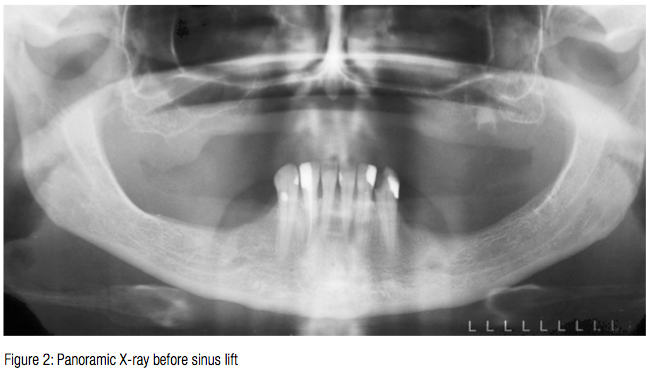
These extractions had contributed to a significant amount of bone loss in the maxilla. The preoperative radiograph (Figure 2) showed that a sinus graft would be necessary in order to place the six implants required for an implant-supported denture. The treatment was defined as a two-stage protocol.

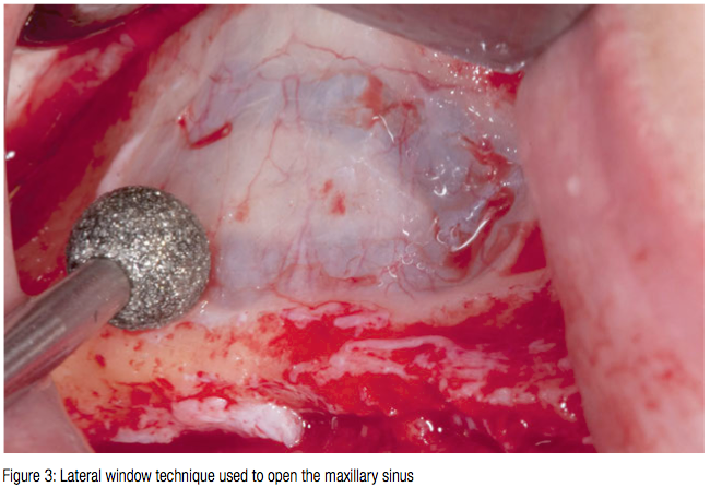
 After discussing the need for general anesthesia, it became evident that the idea of a single operation was very attractive to the patient, and so a single-stage procedure was agreed upon. A lateral window was opened to the sinus (Figure 3). The height of the crestal bone was considered as 1-2 mm maximum. The Schneiderian membrane was lifted without any perforation.

A 3.5 mm diameter osteotomy was prepared in the crestal bone (Figure 4). From the lateral window, a bone ring (Botiss) was then placed in the sinus. A 3.5 mm x 11 mm Ankylos® implant (Dentsply Implants) was inserted from the crest into the sinus through the bone ring (which measured 10 mm in length and 7 mm in diameter) (Figures 5 to 8). The bone ring was gently held in situ with forceps as the implant was inserted.
A cover screw was attached to the implant once it sunk to 11 mm and the insertion adaptor removed. A membrane screw with a pin height of 1 mm was then fixed into the internal threads of the cover screw (Figure 9) until the implant/allograft membrane complex was tight.


The cavity was filled with a small particulated xenograft (Cerabone®, Botiss Biomaterials) (Figure 10). The lateral window was covered with a collagene membrane (Jason®, Botiss Biomaterials ) and fixed with titanium membrane-fixation pins (Ustomed®) (Figure 11). The sinus implants were uncovered after 9 months (Figure 12), and 3 weeks later — after soft tissue healing and impression taking — a removable denture prosthesis was made.
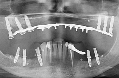
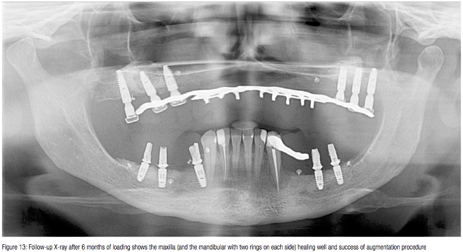
The treatment fulfilled all the patient’s expectations, and in a short time. The follow-up radiograph taken after 6 months of loading shows the perfect healing of the maxilla (Figure 13).

Conclusion
This technique allows for successful bone augmentation with maxillary sinus floor elevation. The authors believe the shortening of the treatment time and the avoidance of a second operation makes the bone ring technique unique in this type of indication. It is possible that unsuccessful sinus lifts where the Schneidarian membrane has been perforated could be dealt with at a single visit too, by using a collagen fleece in the sinus and bone rings without particulate material.
Allogenic graft material
Autologous bone, representing the current “gold standard,” has certain limitations, with the availability of sufficient quantities of autologous bone from intraoral donor sites being restricted. The recent progress in the field of maxillofacial surgery and oral implantology means the need for a predictable and convenient bone grafting material has become increasingly essential.
Although allografts are known to provide a well-established platform for inducing significant osseous regeneration, allogeneic bone tissue appears an adequate alternative. Approximately 40,000 U.S. citizens annually receive allogeneic grafts in the maxillo-mandibular region.
maxgraft® allograft
maxgraft® is exclusively produced from the bone tissue of German, Swiss, and Austrian donors. All pure cancellous bone regeneration material (blocks and granules) originating from living donors are procured from certified procurement centers. All donations from living donors are based on written consent from the patient and highly selective exclusion criteria. Certain risk factors for infectious diseases and internal diseases, as well as current or previous malignancies, are strictly excluded.
Blood samples for serological testing are taken during the explantation of the donor bone tissue, which derives from femoral heads during total hip replacement.
REFERENCES
- Dragoo MR, Irwin RK. A method of procuring cancellous iliac bone utilizing a trephine needle. JPeriodontol. 1972;43(2):82-87.
- Hiatt WH, Schallhorn RG. Intraoral transplants of cancellous bone and marrow in periodontal lesions.JPeriodontol.1973;44(4): 194-208.
- Köndell PA, Mattsson T, Astrand P. Immunological responses to maxillary on-lay allogeneic bone grafts. ClinOralImplantsRes. 1996;7(4): 373-377.
- Schallhorn RG, Hiatt WH. Human allografts of iliac cancellous bone and marrow in periodontal osseous defects. II. Clinical observations. JPeriodontol. 1972;43(2): 67-81.
Stay Relevant With Implant Practice US
Join our email list for CE courses and webinars, articles and mores


