Dr. Anthony Prudenti discusses rehabilitation of a patient with ill-fitting maxillary and mandibular implant-retained overdentures
Abstract
The rehabilitation of failing implant-retained overdentures can be challenging. Working with implants that are placed in a less than ideal position, having a limited amount of prosthetic space, worn-down retentive mechanisms, or old attachments with no replacement parts could render restoring these cases a difficult task. This article describes the use of the new Locator R-Tx® attachment system to rehabilitate a patient with maxillary and mandibular implant-retained overdentures.

Figure 1A: Pretreatment occlusal view of the mandible

Figure 1B: Pretreatment occlusal view of the maxilla
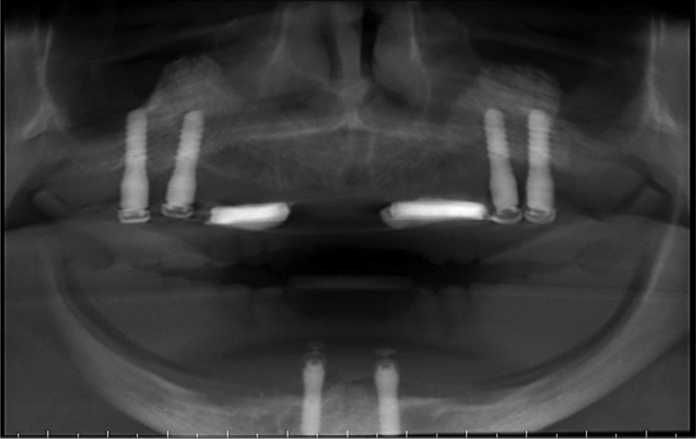
Figure 2A: Pretreatment panoramic radiograph
Introduction
There are functional, psychosocial, and anatomic advantages of implant-retained overdentures for completely edentulous patients.1 There is an enormous amount of evidence in the literature in favor of the implant overdenture retained by two implants in the mandible as the first choice of treatment.2-4 However, there is disagreement on the treatment of choice for the maxillary implant overdenture. There is also no agreement among researchers regarding the ultimate number of implants to be used in the maxilla to retain an overdenture.4-7 However, the distribution and the number of implants have shown to have an effect on the overdenture’s survival.8 Several authors9-12 discussed the importance of having implants that are evenly spaced and distributed in such a way as to provide an increased anterior-posterior spread (A-P spread) and help distribute the load favorably on the implants. Considerations for fulcrum or the axis of rotation created at attachment sites have also been discussed. This article describes the use of the new Locator R-Tx attachment system to rehabilitate a patient who was not satisfied with her existing maxillary and mandibular implant-retained overdentures.

Figures 2B and 2C: Pretreatment radiographs


Figure 3: Occlusal view of the placement of the maxillary anterior implants

Figure 4: Frontal view of the placement of the Locator R-Tx abutments
Clinical report
A highly motivated 67-year-old Caucasian female presented to the clinic for the evaluation of her ill-fitting complete maxillary and mandibular overdentures (Figures 1A and 1B). Her desires were to have complete dentures that were more stable and retentive, while maintaining a palate-less design.
The patient has been a denture wearer since she was in her teens. She was treated a few years ago in an attempt to convert her dentures to implant-retained overdentures. However, complications ensued, leaving her unsatisfied with the dental treatment she received.
Radiographic evaluation and review of the dental history indicated that the patient received bilateral window sinus augmentation and maxillary implant placement in the area of the maxillary right and left first and second molars (Figure 2). Two custom-made cast gold bars, each supported by two implants, retained her maxillary overdenture, while two Locator abutments retained her mandibular overdenture. Following several months of service, both of the gold bars broke due to the large anterior cantilevers, and her maxillary denture was converted to a Locator-retained overdenture. The patient reported that her maxillary overdenture quickly became loose and had been unstable in the largely unsupported anterior region. The maxillary implant distribution that had been designed was insufficient for denture retention and stability. Denture stomatitis of the soft tissue was present under the maxillary denture and was mostly associated with the ill-fitting denture, poor oral hygiene, and the patient not removing her denture. Upon evaluation with the existing dentures in place, the patient exhibited a reduced occlusal vertical dimension, altered speech, and less than ideal esthetics.
The panoramic radiograph demonstrated no obvious pathology. There was evidence of bilateral maxillary sinus augmentation and osseointegrated implants in position of teeth Nos. 2, 3, 14, and 15. Implants were identified as Straumann® tissue level (Straumann®). The four maxillary and two mandibular heavily worn Locator abutments appeared completely seated. Cone beam computed tomography (CBCT) images revealed the knife-edge anterior maxillary ridge with insufficient volume of bone for implant placement.
After discussions with the referring perio-dontist, treatment plans based on clinical findings and radiographic examination were proposed to the patient. She consented to treatment that included maxillary anterior guided bone regeneration (GBR) and placement of implants in positions Nos. 7 and 10, and the fabrication of new maxillary and mandibular implant-retained overdentures.
The surgical phase of therapy completed by the periodontist consisted of guided bone regeneration procedures in the maxillary anterior sextant. A mixture of allograft (Puros®, Zimmer Biomet Dental) and xenograft particulate (Bio-Oss®, Geistlich) with a titanium mesh and collagen membrane was used to perform the graft in an attempt to augment the bone both horizontally and vertically. Following a healing period of 9 months, two OsseoSpeed EV implants 4.8 x 6 mm were placed in the areas of teeth Nos. 7 and 10 (ASTRA TECH Implant System™ EV, Dentsply Sirona) (Figure 3).
Following implant placement and osseointegration, the Locator R-Tx removable attachment system was chosen due to the angulation and orientation of the available maxillary implants. The abutments were selected according to the measured tissue heights and implant diameters, then placed on the implants with the use of a standard hex driver (0.050”/1.25 mm) and torqued as per the manufacturer’s recommendation (Figure 4).
Final impressions were made for the fabrication of the maxillary and mandibular overdentures. The maxillary overdenture was fabricated initially with a full acrylic palate for ease of initial seating. Denture attachment housings were placed on the abutments, and the overdentures were relieved at the abutment-housing sites until passive seating was achieved. The housings were picked up in the dentures intraorally with the use of attachment processing material (CHAIRSIDE®, Zest Dental Solutions). The black processing inserts in both the maxillary and mandibular overdentures were removed and replaced with a combination of gray (zero retention) and blue (low retention) retentive inserts. Then the acrylic in the palate area was removed from the maxillary overdenture. At the 7-week post-insertion visit, the two blue inserts in the mandibular prosthesis were replaced with pink (medium retention) inserts at the patient’s request. Two of three gray inserts in the maxillary prosthesis were replaced with blue inserts, leaving one gray insert in the most off-angled implant at site No. 10, and blue inserts at the remaining five implant sites. Adequate retention and patient comfort were obtained, while allowing the patient’s dexterous ability to properly place and remove the prostheses (Figures 5A-5D).

Figure 5A: Intaglio surface of the maxillary overdenture after the pickup of the denture attachment housings and placement of inserts

Figure 5B: Intaglio surface of the mandibular overdenture after the pickup of the denture attachment housings and placement of inserts

Figures 5C and 5D: Frontal view of patient smile

Discussion
The patient presented with four implants placed too close together in the posterior area of the maxilla, providing a very small A-P spread with a great anterior cantilever. This implant position made the patient’s old dentures unstable and non-retentive because of the unfavorable biomechanics. The patient maintained her desire for a maxillary prosthesis with an open palate. Given the large prosthetic space, an alternative treatment plan would have been the fabrication of custom-milled titanium bars to retain the maxillary overdenture. However, the close proximity of the maxillary implants would necessitate long cantilevers to accommodate the addition of retentive mechanisms to the bar. The patient was deterred from this bar treatment option due to her past negative experience with broken bars and also the associated higher prosthetic costs.
In order to improve the A-P spread and stabilize the prosthesis with a palate-less design, the decision was made to place two implants anteriorly. The guided bone regeneration procedure in the maxillary anterior sextant provided limited available bone volume after bone grafting and dictated the resulting implant positions, which were less than ideal. The buccal inclination of the anterior implants created a divergence between the anterior and the posterior implants, narrowing the choice of individual retentive mechanisms that could be used in order to obtain a path of insertion for the maxillary prosthesis. The Locator R-Tx removable attachment system was selected because of the pivoting capability that allows it to be used with nonparallel implants. In this particular case, given the posterior position of the implants and the patient’s limited dexterity, it would have been difficult for the patient to maintain adequate hygiene with bar attachments, hence the choice of individual retentive mechanisms. In addition, the author believes that the simplicity of this treatment solution will be beneficial for the longevity of the overdenture prosthetic care, along with the simplified patient access for hygiene.
 Anthony Prudenti, DDS, MS, was a Prosthodontics Resident at University of North Carolina at Chapel Hill, School of Dentistry and is currently in private practice in Long Island, New York.
Disclosure: Dr. Prudenti has no financial interests in and is not a consultant for any of the products mentioned in this article.
- Koka S, Baba N, Ma SY. The Benefits of implant overdentures. In: Goodacre CJ, Naylor WP, eds. Implant Overdentures: From Diagnosis to Maintenance. Version 1.0, 2016, published by FOR.org https://www.for.org/. Accessed November 22, 2017.
- Feine JS, Carlsson GE, Awad MA, et al. The McGill Consensus Statement on Overdentures. Montreal, Quebec, Canada. May 24-25, 2002. Int J Prosthodont. 2002;15(4):413-414.
- Thomason JM, Feine J, Exley C, et al. Mandibular two implant-supported overdentures as the first choice standard of care for edentulous patients — the York Consensus Statement. Br Dent J. 2009;207(4):185-186.
- Roccuzzo M, Bonino F, Gaudioso L, Zwahlen M, Meijer HJ. What is the optimal number of implants for removable reconstructions? A systematic review on implant-supported overdentures. Clin Oral Implants Res. 2012;23(suppl 6):229-237.
- Slot W, Raghoebar GM, Vissink A, Meijer HJ. A comparison between 4 and 6 implants in the maxillary posterior region to support an overdenture; 1-year results from a randomized controlled trial. Clin Oral Implants Res. 2014;25(5):560-566.
- Raghoebar GM, Meijer HJ, Slot W, Slater JJ, Vissink A. A systematic review of implant-supported overdentures in the edentulous maxilla, compared to the mandible: how many implants? Eur J Oral Implantol. 2014;7(suppl 2):S191-S201.
- Roccuzzo M, Bonino F, Gaudioso L, Zwahlen M, Meijer HJ. What is the optimal number of implants for removable reconstructions? A systematic review on implant-supported overdentures. Clin Oral Implants Res. 2012;23(suppl 6):229-237.
- Şahin S, Cehreli MC, Yalçın E. The influence of functional forces on the biomechanics of implant-supported prostheses — a review. J Dent. 2002;30(7-8):271-282.
- Mericske-Stern RD, Taylor TD, Belser U. Management of the edentulous patient. Clin Oral Implants Res. 2000;11(suppl 1):108-125.
- Benzing UR, Gall H, Weber H. Biomechanical aspects of two different implant-prosthetic concepts for edentulous maxillae. Int J Oral Maxillofac Implants. 1995;10(2):188-198.
- Kiener P, Oetterli M, Mericske E, Mericske-Stern R. Effectiveness of maxillary overdentures supported by implants: maintenance and prosthetic complications. Int J Prosthodont. 2001;14(2):133-140.
- Lee DJ. Performance of attachments used in implant-supported overdentures: review of trends in the literature. J Periodontal Implant Sci. 2013;43(1):12-17.
Stay Relevant With Implant Practice US
Join our email list for CE courses and webinars, articles and mores

