Drs. Itzhak Binderman, Gideon Hallel, Casap Nardy, Avinoam Yaffe, and Lari Sapoznikov investigate an alternative use for extracted teeth
 Tooth extraction is one of the most widely performed procedures in dentistry, and it has been historically well documented that it can induce significant dimensional changes of the alveolar ridge.
In their review, Horowitz, et al. (2012), stated that less ridge resorption occurs when alveolar ridge preservation procedures are used, compared to leaving fresh alveolar sockets without placing graft material. If performed inadequately, the resulting deformity can be a considerable obstacle to the esthetic, phonetic, and functional results.
In dentistry, allogeneic bone and synthetic mineral materials are the main source for grafting in bone. However, fresh autogenous bone graft is still considered the gold standard since it exhibits bioactive cell instructive matrix properties and is non-immunogenic and non-pathogenic, in spite of the need for harvesting bone and possible morbidity resulting from it.
It is well-known that jawbones, alveolar bone, and teeth develop from cells of the neural crest and that many proteins are common to bone, dentin, and cementum (Donovan, et al., 1993; Qin, et al., 2002). It is, therefore, not surprising that dentin, which comprises more than 85% of tooth structure, can serve as native bone grafting material.

Interestingly, Schmidt-Schultz and Schultz (2005) found that intact growth factors are conserved even in the collagenous extracellular matrix of ancient human bone and teeth.
Methods for processing bovine dentin into particulate and sterile grafting material for preserving of alveolar bone have been described and used in several animal studies (Fugazzotto, et al., 1986; Nampo, et al., 2010; Qin, et al., 2014). It is, therefore, evident that teeth can become grafts that are slowly and gradually replaced by bone (Hasegawa, et al., 2007).
Currently, all extracted teeth are generally considered clinical waste and, therefore, are simply discarded. Recently, however, several studies have reported that extracted teeth from patients, which undergo a process of cleaning, grinding, demineralization, and sterilization, can be a very effective graft to fill alveolar bone defects in the same patient (Kim, et al., 2010; Kim, et al., 2011; Murata, et al., 2011). However, this procedure is extremely time-consuming since the graft is only ready several hours or days after extraction. This article, therefore, aims to present a modified procedure that employs freshly extracted teeth in a clinical setting by re-cycling them into bacteria-free particulate autogenous mineralized dentin for immediate grafting.
A Smart Dentin Grinder® (SDG) (KometaBio) was devised, which grinds and sorts extracted teeth into dentin particulate of a specific size. A chemical cleanser is then applied to process the dentin particulate into a bacteria-free graft over the course of about 15-20 minutes.
This novel procedure is indicated mainly in cases when teeth are extracted because of periodontal reasons and for partially or totally impacted teeth. Teeth that have undergone root canal fillings should not be employed in this procedure because of the risk of contamination by foreign materials. On the other hand, crowns and fillings can be reduced, and the clean dentin of the tooth crown can be processed for immediate grafting.
Method: from extraction to grafting particulate dentin
Teeth without root canal fillings, which have been extracted due to advanced periodontal bone loss or other reasons, such as wisdom teeth extraction or orthodontic indications, can be prepared for immediate grafting.
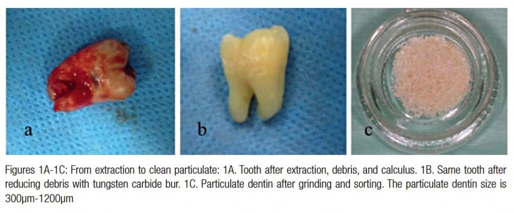
Immediately after extraction, restorations like crowns and fillings should be cut off or removed. Carious lesions and dis-colored dentin, or remnants of periodontal ligament (PDL) and calculus should be reduced by tungsten bur (Figures 1A and 1B). The authors have found that high-speed tungsten carbide burs are most efficient for this process. The roots could be split in case of multi-rooted teeth.
Clean teeth, including crown and root dentin, are dried by air syringe and put into the grinding sterile chamber of the newly designed Smart Dentin Grinder (Figure 2A). The SDG can grind the roots in 3 seconds and then uses the vibrating movement of the grinding chamber to sieve any particles smaller than 1,200µm into a lower chamber that collects particles between 300µm and 1,200µm (Figure 2B). Particles smaller than 300µm fall into a waste drawer, as this fine particulate is not considered to be an efficient size for bone grafting. This grinding and sorting protocol is repeated to grind the remaining teeth particles left in the grinding chamber, still collecting particles between 300µm and 1,200µm.
The particulate dentin from the drawer is immersed in basic alcohol for 10 minutes, in a small sterile glass container. The basic alcohol cleanser consists of 0.5M of NaOH and 30% alcohol (v/v) for defatting, dissolving all organic debris, bacteria, and toxins of the dentin particulate.
 Figure 3 shows the efficiency of the cleanser to dissolve all the organic debris from dentin particulate, including dentin tubules. The scanning electron microscope (SEM) picture shows open and clean tubules after 10 minutes of cleanser treatment (Figure 3C). After decanting the basic alcohol cleanser, the particulate is washed twice in sterile phosphate-buffered saline (PBS). The PBS is decanted, leaving wet particulate dentin ready to graft into freshly extracted sockets, alveolar bone defects, or in procedures involving augmenting the maxillary sinus.
The process from tooth extraction until grafting takes approximately 15-20 minutes.
It should be noted that the efficiency of selecting the dentin particulate of specific size for grafting is more than 95%. It is also obvious that the volume of the particulate dentin is more than twice of the original root volume. Alternatively, the wet particulate can be put on a hot plate (140ºC) for 5 minutes to produce dry, bacteria-free particulate autologous dentin that can serve for immediate or future grafting procedures.
Results: clinical evaluation
 Over a period of 2 years, more than 100 dentists have employed the present procedure for preparing autogenous dentin particulate from extracted teeth for immediate grafting in the same patient. It should be noted that teeth that underwent root canal treatment were discarded. When intact teeth were processed, the enamel and cementum were included. Figures 4 to 7 show a number of typical case presentations where teeth were extracted and processed into bacteria-free particulate autogenous tooth dentin for immediate grafting in same patient.
Wisdom tooth extraction
A total of 16 wisdom teeth, including partially impacted, horizontally impacted, and caries-affected teeth, were processed using the SDG procedure during this study. Figure 4 shows a horizontally impacted LR8 tooth that was in close proximity to the distal root surface of the LR7, creating a deep void. The surgically extracted LR8 exposed the distal root surface of the LR7, almost denuded from bone tissue. The LR8 was processed immediately into the particulate graft, which totally filled the extraction site (Figure 4C). Healing and recovery after the surgical procedure and grafting took place without complications.
A follow-up after 4 months revealed a normal pattern of marginal gingiva around the LR7. Probing was normal: 1 mm-2 mm in depth. On the X-ray distal to the LR7, new bone and particulate dentin was integrated into bone, completely restoring the extraction site and distal bone support of the LR7 (Figure 4D).
Periodontal extractions
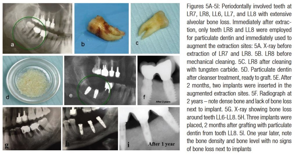
 A further 37 teeth were extracted because of poor periodontal attachment, bone loss, and mobility. Figure 5 illustrates the case of a 56-year-old male patient with a localized, advanced periodontal condition in posterior parts of the mandible.
The LR7 and LR8 were extracted, and the granulation tissue was removed exposing bone tissue walls. The LR7 had a root canal filling and was therefore discarded. The LR8 was processed into particulate dentin by the SDG device and prepared for immediate grafting in the extraction sites.
The grafting of one tooth produced sufficient volume of particulate dentin to overfill the extraction site of both sockets. A Choukroun PRF (platelet rich fibrin) membrane was prepared from the patient’s blood (Cieslik-Bielecka, et al., 2012) to cover the graft. The mucoperiosteum was sutured to the PRF, avoiding tension of tissues. Improved healing was achieved because of the PRF membrane. Approximately 2 months later, two implants were placed, followed by a cemented bridge of LR7-LR8 crowns.
After 2 years, clinical and X-ray follow-up revealed very radiopaque bone integrated into implants, most possibly consisting of bone-dentin but producing a very solid support for implants (Figure 5). A similar procedure was performed in the same patient’s lower left jaw. X-rays showed bone loss around the LL6-LL8 (Figure 5G). Two months after grafting with the particulate dentin from tooth LL8, three implants were inserted (Figure 5H), and 1 year later, the bone density and bone level with no signs of bone resorption at the crest after restoration could be observed (Figure 5J).
Sinus lifts
Autogenous dentin particulate can serve as a superior grafting matrix for augmenting bone in maxillary sinuses, as presented in the next case.
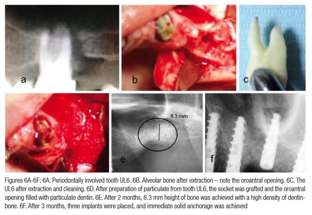
 The patient presented with alveolar bone loss, with infrabony pockets that extended into the maxillary sinus of tooth UL6 (Figure 6). The UL6 was extracted, cleaned, and processed into bacteria-free particulate dentin (Figure 6D). An immediate grafting of the extraction socket was performed, and the tract into the sinus was occluded by the particulate dentin. Closure of the wound and sutures of mucoperiosteum flap was performed.
Healing was normal, and 3 months later, an alveolar ridge of minimum 8.3 mm height was achieved, allowing placement of three implants. It should be noted that one molar — the UL6 — produced at least 2 cc of particulate dentin, which allowed augmentation of the extraction socket and part of the sinus.
Moreover, we found that autogenous dentin grafting allowed the placement of implants after 3 months in the upper jaw because the new bone that was integrated with particulate dentin produced a solid support for implants.
 Loading of implants followed. During preparation of the site for implant placement, a core of bone was recovered from the grafted socket site. The histology revealed new bone integrated with grafted dentin, producing a bone-dentin interface and connectivity (Figure 7).
Discussion
More than 40 years ago, autogenous teeth were routinely transplanted into extraction sockets when possible. It is evident that transplanted teeth that are ankylosed in the jawbone undergo replacement resorption over 5 to 8 years (Sperling, et al., 1986).
In addition, it is well documented that avulsed teeth that are implanted back into their sockets undergo firm reattachment to bone, which is formed directly on root dentin or cementum, leading to ankylosis (Andersson et al, 1989). An ankylosed root is continuously resorbed and replaced by bone, eventually resorbing the entire root, while the alveolar process is preserved during this period and later.
In a recent review, Malmgren (2013) stressed that in ankylosed teeth that are treated by decoronation, the alveolar ridge is maintained in the buccal/palatinal direction, while vertical height is even increased (Park, et al., 2007). Our results reveal similar interaction between the mineralized dentin and osteogenic cells that attach and produce mineralized bone matrix directly on the dentin graft.
A tooth bank in Korea provides a service that prepares autogenic demineralized dentin matrix graft in block or granular types (Kim, et al., 2011; Murata, et al., 2011; Kim, 2012), delaying the grafting procedure from several hours to several days and, therefore, requiring an additional surgical session.
Although demineralized dentin exposes matrix-derived growth and differentiation factors for effective osteogenesis, the newly formed bone and residual demineralized dentin are too weak to support implant anchorage. In contrast, the SDG procedure allows preparation of bacteria-free particulate dentin from freshly extracted autologous teeth, ready to be employed as autogenous graft material immediately.
Mineralized dentin particles have the advantage of maintaining mechanical stability, allowing early loading after grafting in fresh sockets and bone defects. Moreover, in spite of its delayed inductive properties (Yeomans and Urist, 1967; Huggins, et al., 1970), the mineralized dentin is firmly integrated with newly formed bone, creating a solid site for anchorage of dental implants. In fact, our clinical data indicates that implant insertion and loading can be performed in both lower and upper jaws 2 to 3 months after dentin grafting.
Since the mineralized dentin is very slowly remodeled (Yeomans and Urist, 1967; Kim, et al., 2014; Andersson, 2010) in comparison to cortical bone or most biomaterials, the esthetic and structure pattern of the alveolar crest and mucoperiosteum is maintained for years. Teeth and jawbone have a high level of affinity, having a similar chemical structure and composition. Therefore, the authors and others (Kim, et al., 2011; Murata, et al., 2011; Kim, 2012) propose that extracted non-functional teeth or periodontally involved teeth should not be discarded any more.
Extracted teeth can become autogenous dentin, ready to be grafted within 15 minutes after extraction. We consider autogenous dentin as the gold standard graft for socket preservation, bone augmentation in sinuses, or filling bone defects.
Disclosure
The Smart Dentin Grinder is distributed by Kometa Bio. Drs. Itzhak Binderman and Lari Sapoznikov helped to develop the Smart Dentin Grinder and have shares in Kometa Bio Ltd., the company responsible for distributing the device.
Drs. Gideon Hallel, Casap Nardy, and Avinoam Yaffe have no conflict of interest. They participated actively in providing clinical cases and their follow-ups.
References
- Andersson L, Bodin I, Sörensen S. Progression of root resorption following replantation of human teeth after extended extraoral storage. Endod Dent Traumatol. 1989;5(1):38-47.
- Andersson L. Dentin xenografts to experimental bone defects in rabbit tibia are ankylosed and undergo osseous replacement. Dent Traumatol. 2010;26(5):398-402.
- Cieslik-Bielecka A Choukroun J, Odin G, Dohan Ehrenfest DM. L-PRP/ L-PRF in esthetic plastic surgery, regenerative medicine of the skin and chronic wounds. Curr Pharm Biotechnol. 2012;13(7):1266-1277.
- Donovan MG, Dickerson NC, Hellstein JW, Hanson LJ. Autologous calvarial and iliac onlay bone grafts in miniature swine. J Oral Maxillofac Surg. 1993;51(8):898-903.
- Fugazzotto PA, De Paoli S, Benfenati SP. The use of allogenic freeze-dried dentin in the repair of periodontal osseous defects in humans. Quintessence Int. 1986;17(8):461-477.
- Hasegawa T, Suzuki H, Yoshie H, Ohshima H. Influence of extended operation time and of occlusal force on determination of pulpal healing pattern in replanted mouse molars. Cell Tissue Res. 2007;329(2):259-272.
- Horowitz R, Holtzclaw D, Rosen PS. A review on alveolar ridge preservation following tooth extraction. J Evid Based Dent Pract. 2012;12(suppl 3):149-160.
- Huggins C, Wiseman S, Reddi AH. Transformation of fibroblasts by allogeneic and xenogeneic transplants of demineralized tooth and bone. J Exp Med. 1970;132(6):1250-1258.
- Kim YK, Kim SG, Byeon JH, Lee HJ, Um IU, Lim SC, Kim SY. Development of a novel bone grafting material using autogenous teeth. Oral Surg Oral Med Oral Pathol Oral Radiol Endod. 2010;109(4):496-503.
- Kim SG, Kim YK, Lim SC, Kim KW, Um IW. Histomorphometric analysis of bone graft using autogenous tooth bone graft. Implantology. 2011;15:134-141.
- Kim YK. Bone graft material using teeth. J Korean Assoc Oral Maxillofac Surg. 2012;38:134-138.
- Kim YK, Kim SG, Yun PY, Yeo IS, Jin SC, Oh JS, Kim HJ, Yu SK, Lee SY, Kim JS, Um IW, Jeong MA, Kim GW. Autogenous teeth used for bone grafting: a comparison with traditional grafting materials. Oral Surg Oral Med Oral Pathol Oral Radiol. 2014;117(1):e39-45.
- Malmgren B. Ridge preservation/decoronation. J Endod. 2013;39(suppl 3):S67-72.
- Murata M, Akazawa T, Mitsugi M, Um IW, Kim, KW, Kim YK. Human dentin as novel biomaterial for bone regeneration. In: Pignatello R, ed. Biomaterials – Physics and Chemistry. Croatia: InTech; 2011: 127-140.
- Nampo T, Watahiki J, Enomoto A, Taguchi T, Ono M, Nakano H, Yamamoto G, Irie T, Tachikawa T, Maki K. A new method for alveolar bone repair using extracted teeth for the graft material. J Periodontol. 2010;81(9):1264-1272.
- Park CH, Abramson ZR, Taba M Jr, Jin Q, Chang J, Kreider JM, Goldstein SA, Giannobile WV. Three-dimensional micro-computed tomographic imaging of alveolar bone in experimental bone loss or repair. J Periodontol. 2007;78(2):273-281.
- Qin C, Brunn JC, Cadena E, Ridall A, Tsujigiwa H, Nagatsuka H, Nagai N, Butler WT. The expression of dentin sialophosphoprotein gene in bone. J Dent Res. 2002;81(6):392-394
- Qin X, Raj RM, Liao XF, Shi W, Ma B, Gong SQ, Chen WM, Zhou B. Using rigidly fixed autogenous tooth graft to repair bone defect: an animal model. Dent Traumatol. 2014;30(5):380-384.
- Schmidt-Schultz TH, Schultz M. Intact growth factors are conserved in the extracellular matrix of ancient human bone and teeth: a storehouse for the study of human evolution in health and disease. Biol Chem. 2005;386(8):767-776.
- Sperling I, Itzkowitz D, Kaufman A, Binderman I. A new treatment of heterotransplanted teeth to prevent progression of root resorption. Endod Dent Traumatol. 1986;2(3):117-120.
- Yeomans JD, Urist MR. Bone induction by decalcified dentin implanted into oral, osseous and muscle tissues. Arch Oral Biol. 1967;12(8):999-1008. 46 Implant practice Volume 8 Number 2
Stay Relevant With Implant Practice US
Join our email list for CE courses and webinars, articles and mores


