
Educational aims and objectives
This self-instructional course for dentists aims to discuss the Posterior Lateral Mandibular Cortical Stabilization (PLMCS) and Posterior Medial Mandibular Cortical Stabilization (PMMCS) techniques for implant placement.
Expected outcomes
Implant Practice US subscribers can answer the CE questions by taking the quiz online to earn 2 hours of CE from reading this article. Correctly answering the questions will demonstrate the reader can:
- Realize some advantages of PLMCS and PMMCS.
- Realize some of the disadvantages of PLMCS and PMMCS.
- Recognize some clinical issues that can arise during these procedures.
- Identify some safety measures that can be taken when using these techniques.
In this CE, Dr. Sami Nizam illustrates how the full-arch implant placement techniques of PLMCS and PMMCS can provide successful long term outcomes even in the most atrophic cases. Subscribers who pass the quiz can receive 2 CE credits!
Dr. Sami Nizam discusses techniques for full-arch implant placement
Overview
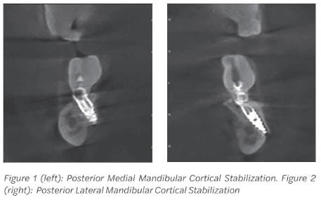
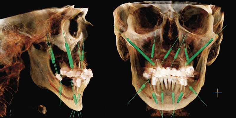
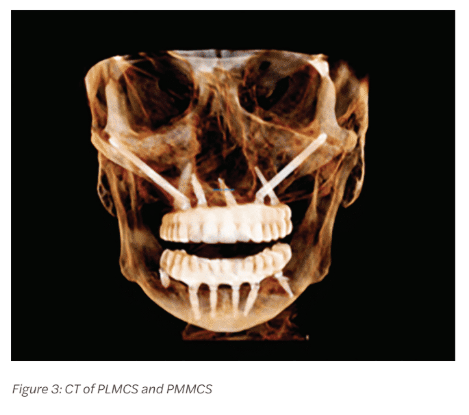
Full arch implant placement is becoming increasingly common. Techniques have advanced significantly even over the past 10 years. Most of these advancements have focused onatrophic cases of the maxilla allowing restoration of full dentition without cantilevers. These same advancements have lagged for atrophic mandibles. Posterior Lateral Mandibular Cortical Stabilization (PLMCS) and Posterior Medial Mandibular Cortical Stabilization (PMMCS) are techniques that were developed for extending AP spread, cantilever elimination, and increasing insertional torque in the posterior mandibular region. They do this by engaging the lateral or medial posterior mandibular cortex (Figures 1-3) regions that are present even on severely atrophic cases, while avoiding the inferior alveolar nerve. As bone in these cortices is type I, excellent immediate stability can be achieved primarily. This allows reliably high composite torque scores and immediate loading of the temporary prothesis.
Surgical technique
One of the principle advantages of this technique is it follows the same principles of angled implant placement that any full-arch surgeon is comfortable with. In the author’s practice, full-arch cases are performed under general intubated anesthesia. Placement begins with a standard full thickness mucoperiosteal flap elevation. The flap is continued distally with a buccal release for PLMCS or tapering slightly lingual for PMMCS. One must be aware that the lingual nerve extends to the level of the ridge or above in up to 17% of cases, in some studies, and the incision should never track past the lingual cortex.1 Dissection is taken to the buccal or lingual in a subperiosteal fashion depending on the technique chosen. This is particularly important using PMMCS as violation of the soft tissue will result in prolapse of the sublingual or submandibular gland. This can be an intraoperative annoyance and lead to a sialocele blocking the duct over the long term.
The author usually makes the decision on which technique to use based on which direction gives better implant-to-bone contact area. Cadaveric dissections have revealed the inferior alveolar nerve travels from medial to lateral in a horizontal plane as it traverses the body of the mandible anteriorly.2 Due to this typical trajectory, PLMCS is selected approximately two-thirds of the time. This allows a longer/wider implant with resultant greater bone-to-implant contact area. Retraction of the soft tissue is typically with Minnesota retractor on the buccal. For the lingual, the tongue is retracted with a Sweetheart (SurgiMac) and soft tissues subperiosteally with a Seldin or similar retractor. This retraction can be challenging in a poorly sedated patient and cause airway embarrassment (airway compromise) from posterior tongue pressure.
Next, the implant preparation is begun with a pilot bur visualizing the apex of the bur exiting out the cortex. This is much like a zygomatic implant. For added safety with this technique, viewing the proposed entry and exit point on a coronal CT and using measurements if desired can help ensure the nerve is not penetrated. As is common in the posterior mandible in atrophic cases, the marrow space is of type 3 or 4 bone. Densification burs can be used to compact the bone. They give the added benefit of guiding over the canal as it is surrounded in type 1 or 2 bone.
An additional safety measure one could take, if the clinician desires, would be active or static navigation. The implant is then placed and countersunk. A bone mill is used to remove
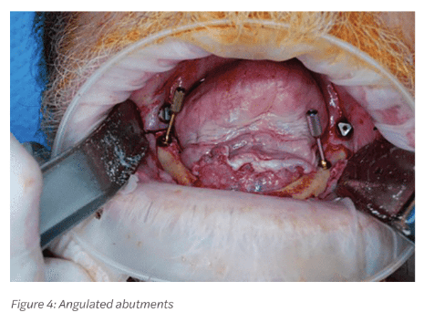
excess bone around the coronal portion. If the tip is overextended, it can be cut with copious irrigation, again much like a zygomatic implant. Caution must be exercised so that the bur does not catch the lingual or buccal tissue as significant tissue damage can occur. Copious irrigation is also obligatory as heat generation can be significant. Typically a 30 degree multiunit is then placed and torqued to specification (Figure 4). On occasion, a more acute angle multiunit abutment may be needed for the final restoration, as the max divergence for monolithic zirconia is no more than 25-30 degrees. If this cannot be achieved, then a custom multiunit (or 45 degree multiunit if available for the brand being used) may be necessary. The author has had this issue occur once, and it was resolved with a custom multiunit. Attached gingiva can be sparse in this region.
Literature and clinical experience have revealed a lack of attached gingiva can lead to peri-implantitis, recession, and bone loss.3 A plan for a free gingival graft with the goal of having 2 mm or greater of keratinized gingiva should be considered once integration has occurred. The author has found this the best time to place free gingival grafts as the temporary can be used as an effective healing barrier with simple composite addition. One can also evaluate the amount of tissue needed, demonstrate the deficit to the patient with intraoral photography, and plan if other sites will be needed before executing the procedure.
Advantages
Protocols such as PATZI (Pterygoid, Angled Anterior, Transnasal, Zygomatic) have revolutionized the treatment of the atrophic maxilla.4 Benefits have been clear. Using PLMCS and
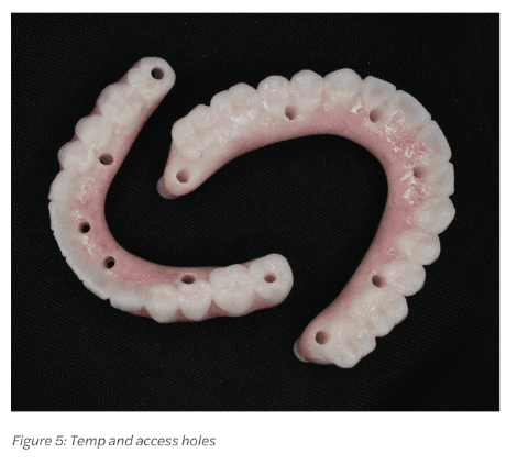
PMMCS in the mandible brings many of the same benefits including greater composite torque scores, larger AP spreads, ability to load, and the ability to restore to second molar even in the temporary (Figure 5). It is the author’s opinion that patients should receive second molars when feasible in a temporary. Distributing forces over a larger area decreases force at any point. Remembering the law of physics, pressure=force/area. As area (the denominator) in the formula increases, with force (the numerator) held constant, pressure then decreases. This is very similar to the concept of a bed of nails. One cannot lie on a single nail; however, one can lie on a bed of nails without harm. This concept has been born out in the literature. Decreasing distal cantilevers causes less stress on the prosthesis distal junction as well as less crestal forces on the distal implant.5 Although there is no exact agreement on cantilever length, it is accepted it should never be greater than two times the AP spread with 1.5 being most commonly used in practice.6 PLMCS and PMMCS allow complete elimination of any cantilever.
Other challenging clinic scenarios are patients who are Class III, who have anterior mental foramen, or U-shaped mandibles. These scenarios can limit the ability to replace a second molar and sometimes even a full first molar if implants are placed anterior to the foramen using standard All-on-4 procedures. Figures 6 and 7 demonstrate such a scenario. The patient has a Class I occlusion, usual anatomic exit of the mental foramen, however, has a U-shaped mandible. This causes a small AP spread of only 7 mm in this case. The average
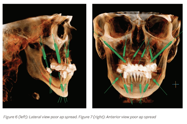 male Caucasian molar is approximately 11.5 mm in MD width.7 In this scenario, one molar would be pushing the AP spread limits, and two would be prohibitive. This logically does not make sense when we have the ability to provide a second molar on the maxilla using PATZI protocols.
Another clinical problem that occurs is what to do in cases of traditional All-on-4 with a distal tilted implant anterior foramen that fails. Depending on when it occurs and the amount of bone loss, adequate bone stock may not be available to place another implant, particularly immediately. PLMCS and PMMCS provide a “bail out” to keep the patient in a fixed prosthesis in these scenarios. There also is the theoretical advantage of applying forces into the body of an atrophic mandible to avoid atrophy in this region. The author has seen fractures occur in this region from falls on patients with traditional All-on-4s. These techniques have the potential to mitigate resorption in this region.
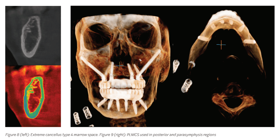
The body region of the mandible is often of type 3 or 4 bone in edentulous cases. Figure 8 demonstrates a case in which extreme type 4 bone was present in the body extending to the parasymphysis region. Traditional implants with densification burs yielded no primary stability in the angled or posterior implant. Engaging type 1 bone of the cortex buccally in these regions allowed for excellent primary stability and case loading seen in Figure 9. Finally, practice differentiation is key in today’s market. The ability to fully restore dentition immediately, reliably, and with long term success can be a strong practice builder and marketing tool.
Disadvantages
As with any increased AP spread and arch length impression, accuracy can be affected.8 Accuracy of digital or traditional impression techniques becomes more important to avoid misfits. Additionally, there is the theoretical disadvantage of mandibular flexure and long spans causing fracture in the temp or final.9 It is recommended that if PLMCS or PMMCS is used, it be used with four anterior implants for better biomechanical resistance. In severely atrophic mandibles, fracture could be a theoretical disadvantage, especially in the case of a tapered implant that is overtorqued during placement. This is fortunately unlikely as the opposite cortex as well as the inferior border remains intact.
Milling difficulties can occur in the final, particularly monolithic zirconia. This can occur if the implant is placed at too great of an angle and cannot be up righted with a multiunit abutment. In this case, a custom multiunit abutment would need to be fabricated. Inferior alveolar nerve injury is also a potential sequela. Techniques listed above such as visualization, measurement on the CT, feel, and densification burs are usually adequate. If one prefers, active or static navigation techniques could be implemented to further mitigate risk.
Injury to soft tissue on the buccal could theoretically result in injury to the facial artery or vein as they cross the inferior border of the mandible. If this occurs, localization and ligation would be required. Injury to the lingual tissue could result in injury to the lingual nerve, submandibular gland, or sublingual gland depending on region of placement. Glandular injury could lead to sialocele. Conservative injection of neuromodulator at the time of injury, if witnessed, would offer a simple solution. If a sialocele is noted late, marsupialization would offer the simplest solution. As with any complications, the easiest solution is avoidance, and good retraction with the patient asleep is a prerequisite for these techniques. The author has experienced none of the above complications to-date.
A final disadvantage is adding of cost and time in comparison to a traditional All-on-4. This should be factored into the cost presented to the patient. The author has found that when the advantages are discussed using CTs and printed temporaries, it becomes a great selling point.
As full-arch implant placement becomes more mainstream, practice differentiation becomes crucial. PLMCS and PMMCS give the ability to restore a full dentition in the temporary, increase composite torque scores, eliminate cantilevers, better distribute occlusal forces, and provide better long term outcomes even in the most atrophic cases. It is the author’s sincere opinion that this technique is a valuable tool to anyone involved in the practice of restoring full arches.
For more techniques in full-arch implant placement, read Dr. Leke Olowokere’s “Primary stability — an overview of options for the full-arch implantologist,” here: https://implantpracticeus.com/?s=full+arch+implant+placement.
References
- Nizam SA 2nd, Ziccardi VB. Trigeminal Nerve Injuries: Avoidance and Management of Iatrogenic Injury. Oral Maxillofac Surg Clin North Am. 2015 Aug;27(3):411-424. doi: 10.1016/j.coms.2015.04.006. Epub 2015 Jun 2.
- Yeh AYE, Finn BP, Jones RHB, Goss AN. The variable position of the inferior alveolar nerve (IAN) in the mandibular ramus: a computed tomography (CT) study. Surg Radiol Anat. 2018 Jun;40(6):653-665. doi: 10.1007/s00276-018-1973-9. Epub 2018 Jan 20.
- Ramanauskaite A, Schwarz F, Sader R. Influence of width of keratinized tissue on the prevalence of peri-implant diseases: A systematic review and meta-analysis. Clin Oral Implants Res. 2022 Jun;33 Suppl 23:8-31. doi: 10.1111/clr.13766.
- Ponnusamy S, Gonzalez J, Holtzclaw D. A Systematic Approach to Restoring Full Arch Length with Maxillary Fixed Implant Reconstruction: The PATZi Protocol. Int J Oral Maxillofac Implants. 2023 Oct 17;38(5):996-1004. doi: 10.11607/jomi.10153.
- Khorshid HE, Issa NO, Ekram AM. Effect of implant diameter and cantilever length on the marginal bone height changes and stability of implants supporting screw retained prostheses: A randomized double blinded control trial. J Adv Prosthodont. 2023 Jun;15(3):101-113. doi: 10.4047/jap.2023.15.3.101. Epub 2023 Jun 28.
- Wang Q, Zhang ZZ, Bai SZ, Zhang SF. Biomechanical analysis of stress around the tilted implants with different cantilever lengths in all-on-4 concept. BMC Oral Health. 2022 Nov 5;22(1):469. doi: 10.1186/s12903-022-02520-8. PMID: 36335327;
- Singh SP, Goyal A. Mesiodistal crown dimensions of the permanent dentition in North Indian children. J Indian Soc Pedod Prev Dent. 2006 Dec;24(4):192-196. doi: 10.4103/0970-4388.28076.
- Zhang YJ, Qian SJ, Lai HC, Shi JY. Accuracy of photogrammetric imaging versus conventional impressions for complete arch implant-supported fixed dental prostheses: A comparative clinical study. J Prosthet Dent. 2023 Aug;130(2):212-218. doi: 10.1016/j.prosdent.2021.09.035. Epub 2021 Nov 12.
- Gao J, Li X, He J, Jiang L, Zhao B. The effect of mandibular flexure on the design of implant-supported fixed restorations of different facial types under two loading conditions by three-dimensional finite element analysis. Front Bioeng Biotechnol. 2022 Aug 29;10:928656. doi: 10.3389/fbioe.2022.928656.
Stay Relevant With Implant Practice US
Join our email list for CE courses and webinars, articles and mores