Educational aims and objectives
This article aims to describe the essential guidelines for using CBCT in implant dentistry.
Expected outcomes
Implant Practice US subscribers can answer the CE questions by taking the quiz to earn 2 hours of CE from reading this article. Correctly answering the questions will demonstrate the reader can:
- Identify radiation dose and optimization of exposure.
- Recognize the biological risks of irradiation.
- Realize radiation protection and the core standards for safe and effective use of CBCT technology.
- Identify the ethical and medicolegal considerations pertaining to the clinical application of CBCT scanners in implant dentistry.
- Realize how to maximize the diagnostic and treatment planning benefits of CBCT to best serve the interests of the patient, optimize patient safety, and minimize radiation-related patient risk.
Dr. Johan Hartshorne finished his 4-part series on radiation from CBCT.
Dr. Johan Hartshorne assesses the clinician liability and patient risk from radiation when using CBCT imaging by looking at the potential pitfalls and limitations
Introduction
The role of 3D cone beam computed tomography (CBCT) imaging as a new diagnostic tool in modern-day dentistry cannot be overemphasized and has increasingly been referred to as the new professional “standard of care” for diagnostic maxillofacial imaging (Curley and Hatcher, 2009; Tipton and Metz, 2008; Zinman, et al., 2010).
It serves as an essential diagnostic tool for clinical assessment and treatment planning and has revolutionized every aspect of how dental implant practices are performed (Hatcher, et al., 2003; Kobayashi, et al., 2004; Sato, et al., 2004). However, CBCT technology does not come without pitfalls, liabilities, and risks.
Understanding the radiation dose imparted by CBCT and potential biological risks to the patient is an important patient safety issue. Appropriate selection criteria must be used with the minimum radiation exposures that result in images of acceptable diagnostic qualities (Carter, et al., 2008; Horner, et al., 2009).
As with any new technology introduced to a profession, the education lags far behind the technological advance; this is especially true of cone beam imaging. An important basic requirement of using CBCT imaging as a diagnostic and treatment planning tool is that practitioners should have appropriate training and competencies to ensure safe and effective use of a CBCT unit that will best serve the patient’s interests, while optimizing patient safety and minimizing radiation-related patient risk.
Purpose
The purpose of this article (the fourth and final part of this series) is to provide an overview of the following topics:
- Radiation dose and optimization of exposure
- The biological risks of irradiation
- Radiation protection and the core standards for safe and effective use of CBCT technology
- The ethical and medicolegal considerations pertaining to the clinical application of CBCT scanners in implant dentistry
This knowledge will enhance a practitioner’s understanding of how to maximize the diagnostic and treatment planning benefits of this technology that best serve the interests of the patient, while optimizing patient safety and minimizing radiation-related patient risk.
Radiation dose, risks, safety considerations, and optimization of exposure
Radiation dose and optimization of exposure
Among the many risks to which we are prone is the normal background radiation with a world average of about 2.4mSv per individual and year. Medical exposures now contribute to around 20% of the average annual per head effective dose of the global population. Medical diagnostic X-ray examinations result in per head effective dose of 0.6mSv of which dental radiology contributes only a small fraction (0.01mSv) (Harris, et al., 2012; United Nations Scientific Committee on the Effects of Atomic Radiation, 2010).
Understanding the radiation dose imparted to the patient by dental radiology is an important safety issue. Appropriate selection criteria must be used with the minimum radiation exposures that result in images of acceptable diagnostic quality (Figure 1) (Carter, et al., 2008; Horner, et al., 2009). This concept is known as “as low as reasonably achievable” (ALARA) (Figure 2) (Farman, 2005; Tyndall, et al., 2012).

In general, imaging parameters (such as kV, mAs, and field of view [FOV] size) have an effect on the effective radiation dose, as well as image quality parameters (spatial resolution, contrast, noise, and artifacts) (Pauwels, et al., 2012; 2015). In terms of optimization of exposure, the most straightforward imaging parameter is FOV size, as larger FOVs increase radiation dose to the patient. Effective doses for different CBCT devices exhibit a wide range, but for all devices, significant dose reduction can be achieved by reducing the FOV to the actual region of interest (Bornstein, et al., 2014). In addition, larger FOVs increase the relative amount of scattered radiation reaching the detector, leading to an increase in noise and artifacts, thus affecting the quality of the image. Therefore, FOVs should always be kept as small as possible, covering only the area of interest (SEDENTEXCT, 2011).
Increased radiation dose risk
Patient risk and clinician liability from radiation has been a continuing concern in oral and maxillofacial imaging, due to the frequency of radiographic examinations in dental practice (Friedland and Miles, 2014).
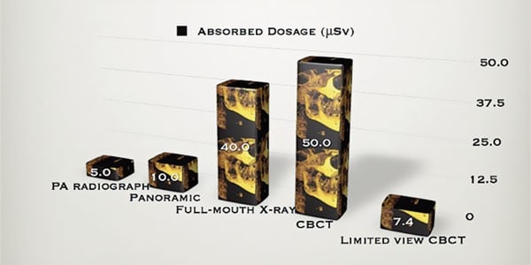
With the increased use of CBCT imaging in dental practice, clinicians must be made aware that patient radiation doses associated with CBCT imaging are higher than those of conventional radiographic techniques (Figure 1). Therefore, routine replacement of current radiographic techniques must be considered with great care (Bornstein, et al., 2014).
The average effective radiation dose for a CBCT corresponding to a small FOV is 34µSv, medium FOV is 88µSv, and a large FOV is 131µSv with the medium and large FOVs showing the largest variability of doses (Pauwels et al, 2012). In general, CBCT imaging results in higher patient doses than standard radiographic methods used in dental practice for dental therapy, but remain well below those reported for common multidetector computed tomography protocols (280µSv to 1410µSv) (Bornstein, et al., 2014; SEDENTEXCT, 2011).
The effective radiation dose for a CBCT is 2- to 4-times greater than for a cephalometric X-ray (<6µSv); 3- to 6-times greater than a panoramic X-ray (2.7µSv to 24.3µSv); and 9- to 14-times greater than a periapical X-ray (<1.5µSv) (Bornstein, et al., 2014; SEDENTEXCT, 2011). Risks have also been noted in the radiation dose needed with CBCT, although it is generally believed the radiation dose of CBCT is significantly lower than a conventional CT (Schulze, et al., 2004).
Effective radiation doses of typical dentomaxillofacial applications are relatively low when compared with the worldwide annual background radiation of 200µSv to 4500µSv (Green, et al., 1992; Jacobs and Quirynen, 2014). The effective radiation dose of CBCT can be affected to an order of magnitude by patient size, FOV, region of interest, and resolution. A careful selection of all these parameters is needed to optimize diagnostic information and reduce the patient’s radiation exposure (Figure 2) (European Academy of Dental and Maxillofacial Radiology, 2012).
Biological risks of irradiation
The biologic effects of ionizing radiation maybe divided into two categories: tissue reactions (previously called deterministic effects) and chromosomal effects (also known as stochastic effects) (International Commission on Radiological Protection, 2007). Tissue reactions are proportional to the dose and occur in all individuals when the dose is large enough. They result in cell death or cell malfunction, and the severity of the effects increases with the dose. Tissue reactions — such as cataract formation, skin erythema, and effects — on fertility occur only above threshold doses that are far greater than those given in dental radiology. Chromosomal effects can be considered as “chance” (or stochastic) effects, where the magnitude of the risk is proportional to the radiation dose (SEDENTEXCT, 2011).
Chromosomal or stochastic effects include the detriment-adjusted nominal risk of cancer and hereditable effects owing to mutation of reproductive cells. The detriment-adjusted risk factor for the whole population is 5% per Sv. In case of cancer, epidemiological and experimental studies provide evidence of radiation risk, albeit with uncertainties at low doses (<100mSv).
The probabilistic nature of stochastic effects makes the distinction between “safe” and “harmful” exposures to radiation impossible (International Commission on Radiological Protection, 2007; SEDENTEXCT, 2011). The biological risk from irradiation is age-dependent, being highest for the young and least for the elderly (SEDENTEXCT, 2011). The risk for small children is 3 times the risk for an adult at 30 years of age.
Radiation protection — limiting the dose and risk from X-ray imaging
Radiation protection in clinical practice is based on two fundamental principles (International Commission on Radiological Protection, 2007). The first principle is that of justification. The clinician has an obligation to ensure that there must be a net benefit for the individual who is being exposed; for example, potential benefits must outweigh the potential risks (Harris, et al., 2012).
The second principle is that of optimization of radiation exposure; namely, it should be as low as reasonably achievable to minimize the risk of cancer or tissue effects (Figure 2) (Harris, et al., 2012; International Commission on Radiological Protection, 1977). It is obvious that CBCT should not be carried out without proper optimization strategies to maintain the correct balance between cost and radiation dose on the one hand, and information required, on the other hand. Therefore, the scanned area should not exceed the area of interest. This would substantially limit the dose of radiation, while justifying the use of CBCT in preparing for implant surgery (Jacobs and Quirynen, 2014).
The FOV of the CBCT examination should be restricted to the region of interest (ROI) whenever possible. Patient and equipment-specific dose reduction measures should be used at all times (Bornstein, et al., 2014).
Basic principles and core standards for safe and effective practice
The clinician performing or interpreting CBCT scans for implant dentistry should take into consideration current radiologic guidelines for safe and effective use of CBCT. The European Association of Dental and Maxillofacial Radiology developed the following consensus-based core standards for safe and effective use of dental CBCT (Horner, et al., 2009). These basic principles are not in conflict with the current evidence-based guidelines as set by SEDENTEXCT, a collaborative project of the European Union (SEDENTEXCT, 2011).
Justification
- CBCT examinations must not be carried out unless a history and clinical examination has been performed.
- CBCT examinations must be justified for each patient to demonstrate that the benefits outweigh the risks.
- CBCT examinations should potentially add new information to aid the patient’s treatment management.
- CBCT should not be repeated routinely on a patient without a new risk/benefit assessment having been performed.
- When accepting referrals from other dentists for CBCT examinations, the referring dentist must supply sufficient clinical information to allow the CBCT practitioner to perform the justification process.
- CBCT should only be used when conventional (lower dose) radiography does not provide adequate information for the question at stake.
- CBCT images must undergo a thorough clinical evaluation of the entire image data set.
- Where it is likely that the evaluation of the soft tissues will be required as part of the patient’s radiological assessment, the appropriate imaging should then be conventional CT or MR, rather than CBCT.
Optimization
- CBCT examinations must use the smallest volume size (FOV that is compatible with the clinical situation).
- Where CBCT equipment offers a choice of resolution, the resolution compatible with adequate diagnosis and the lowest achievable dose should be used.
- A quality assurance program must be established and implemented for each CBCT facility, including equipment, techniques, and quality control procedures.
- Aids to accurate positioning, such as light beam markers, must always be used.
Quality standards and assurance
- All new installations of CBCT equipment should undergo a critical assessment and detailed acceptance tests before use to ensure that radiation protection for staff, members of public, and patients are optimal.
- CBCT equipment should undergo regular routine tests to ensure that radiation protection for both practice/facility users and patients has not deteriorated.
Staff protection
- For staff protection from CBCT equipment, the guidelines in Section 6 of the European Commission document, European guidelines on radiation protection in dental radiology should be followed (European Commission, 2004).
- A qualified expert should oversee the installation and use of CBCT to ensure staff doses are ALARA and all relevant national requirements are met (SEDENTEXCT, 2011).
- CBCT equipment should be installed in a protected enclosure and the entire enclosure designated a controlled area. The provision of personal monitoring should be considered (SEDENTEXCT, 2011).
Training and competence
- All those involved with CBCT must have received adequate theoretical and practical training for the purpose of radiological practices and relevant competence in radiation protection.
- Continuing education and training after qualification are required, particularly when new CBCT equipment or techniques are adopted.
- Dentists responsible for CBCT facilities who have not previously received adequate theoretical or practical training should undergo a period of additional theoretical and practical training that has been validated by an accredited academic institution.
- For dentoalveolar CBCT images of teeth, their supporting structures, the mandible and the maxilla up to the floor of the nose (such as 8 cm x 8 cm FOV), clinical evaluation should be made by a specially trained dentomaxillofacial radiologist or where this is impracticable, an adequately trained general dental practitioner.
- For non-dentoalveolar small FOV (such as temporal bone) and all craniofacial CBCT images (FOV extending beyond the teeth, their supporting structures, the mandible, including temporomandibular joint disorders and the maxilla up to the floor of the nose), clinical evaluation should be made by a specially trained dentomaxillofacial radiologist or by a medical radiologist.
CBCT: Ethical and medicolegal considerations
As CBCT technology advances in dentistry, clinicians who embrace it should understand the potential liabilities and risks associated with the technology, as well their ethical obligations toward the patient and profession.
Competency and serving the best interest of the patient
Given that a single CBCT scan uses ionizing radiation at levels exceeding any 2D dental-imaging modalities, it is timely to recommend the development of rigorous training standards in maxillofacial CBCT imaging in the interests of our patients who deserve to have imaging performed by competent clinicians (Scarfe, et al., 2006).
Furthermore, there is mounting concern among oral and maxillofacial radiologists, based on issues of quality and patient safety, that dentists with inadequate training and experience should not perform interpretation of extended FOV diagnostic imaging studies using CBCT (Scarfe, et al., 2006). The view has also been expressed by the American Association of Oral and Maxillofacial Radiology that non-radiologist dentists should not be excluded from performing CBCT imaging, provided they have appropriate and documented training and experience (Scarfe, et al., 2006).
If dentists or non-radiologist dental specialists decide to examine their own volumes, then they are held to the same standard of care as an oral maxillofacial radiologist and required to find and report any unusual conditions or incidental findings that may reside in that volume. This is a significant responsibility, one that might require additional training for some dentists (Miles and Danforth, 2014).
Unfortunately, many dentists who acquire a CBCT unit for their practice are not adequately trained to fully understand and take advantage of its capabilities. Common errors include a scan with an FOV that is too large or too small. A larger FOV than is necessary has at least two deleterious effects. It results in a less optimal image (the larger the FOV, the greater the scatter and the less detailed the image) and a greater patient dose.
Although increased scatter and dose are not likely to result in liability, a wider FOV than is necessary does have medicolegal implications as far as interpretation of the image is concerned (Friedland and Miles, 2014). The potential liability issue that arises is that the dentist is responsible for reading the entire scan. Thus, even though the maxilla and skull were not required in this case, once they are included in the scan, the dentist is responsible for pathology missed in either location (Friedland and Miles, 2014).
Practitioners should have appropriate training in operating a CBCT unit and competence in interpreting images. This training and competence should be maintained through continuing dental education courses. Such training should include a thorough review of normal maxillofacial anatomy, common anatomic variants, and imaging signs of diseases and abnormalities. This is particularly important for CT and CBCT imaging because of the complexity of structures within the expanded FOVs (Carter, et al., 2008).
Clinicians who do not have adequate experience in interpreting a CBCT scan should rather refer the scan to a specialist radiologist or radiographer for reviewing, or refer the patient to a specialist oral and maxillofacial radiologists for the CBCT scan and reviewing.
Ethical obligation of beneficence
Dentistry exposes practitioners at each patient encounter to an ethical obligation of beneficence; namely, will the image serve the patient’s best interests?
Practitioners are obliged ethically and morally to measure the benefit from using a CBCT scan versus the potential risk from radiation before a decision is made to take a CBCT scan (SEDENTEXCT, 2011). Dentists may also have legal responsibilities and duties that overlap with ethical considerations to some degree (Scarfe, 2011).
When a patient undergoes an X-ray examination, millions of photons pass through their body. These can damage any molecules by ionization, but damage to DNA in the chromosome, although rare, is of particular importance because a chromosome can be permanently altered in the process, ultimately leading to the formation of a tumor (SEDENTEXCT, 2011). While doses and risks for dental radiology are small, a number of epidemiological studies have provided some limited evidence of an increased risk of brain (Longstreth, et al., 1993; Claus, et al., 2012), salivary gland (Horn-Ross, et al., 1999), and thyroid tumors (Memron, et al., 2010) for dental radiographs. Any radiation exposure entails a risk to the patient. Under normal circumstances, however, the risk from dental radiography is very low.
The basic premise that justifies the use of CBCT is that the benefits provided by the radiographic examination must far outweigh the potential risks associated with radiation exposure (SEDENTEXCT, 2011). Such benefits may be in the form of increased diagnostic efficacy, enhanced treatment planning, or better therapeutic outcomes (Harris, et al., 2012; Mallya, 2015).The clinician has an obligation to ensure appropriate measures are taken at all instances to limit radiation dose and risk to the patient through justification and optimization processes. A CBCT examination should, at all times, add new information to aid the patient’s treatment (SEDENTEXCT, 2011). The ALARA principle must also be applied, taking into account any alternative techniques that might achieve the same objectives (Harris, et al., 2012).
Informed patient consent
Reporting the findings in a CBCT volume is probably the most essential process in the total diagnostic evaluation of a patient, even if it is something as simple as implant planning (Miles and Danforth, 2014).
It is the responsibility of healthcare providers to acquire information data from patients to best determine the health status of patients, and if treatment is indicated, to provide a basis for treatment planning and informed patient consent (Miles and Danforth, 2014). The CBCT report, therefore, becomes an important component of information for patients to make informed decisions about their health or dental care and treatment, and to facilitate giving informed consent standard of care.
A CBCT could give the clinician the opportunity to visualize, plan, and explain the situation to the patient more clearly. Hence, the standard of care by definition involves that, in all the cases where it is required and possible, the CBCT or some 3D technique should be offered to the patient. In the case that the patient declines after being informed of the risks, benefits, and alternatives, then informed refusal should be obtained and documented (Curley and Hatcher, 2009).
Medicolegal issues on reviewing and reporting
The interpretation of CBCT images remains one of the most vexing problems and greatest source of liability for practitioners who take their own scans (Friedland and Miles, 2014). The use of CBCT carries with it medicolegal risks of which the dental practitioner should be aware. These include licensing and malpractice liability concerns (Friedland and Miles, 2014).
The quality, accuracy, and use of the CBCT report or findings are subject to medicolegal scrutiny, and knowledge of such issues determines whether or not a primary provider or a secondary radiology reader evaluates the image data and issues the final report (Miles and Danforth, 2014). If dental practitioners own a CBCT machine and take scans not only for their own patients, but also for those referred from outside, they must ensure in the latter situation that they do not lack malpractice coverage in the event of a lawsuit.
Some malpractice carriers have explicit limitations and will defend a dental practitioner for CBCT-related diagnostic issues only if the suit is brought by one of the dentist’s own patients. If a patient who was referred from outside solely for a CBCT scan brings a lawsuit for a misdiagnosis, then malpractice coverage can be denied (Friedland and Miles, 2014).
When examining a CBCT scan, the dental practitioner has the legal responsibility to examine every cross section to ensure that no pathosis is overlooked. The accepted standard of care is that the dentist reviewing/reading the CBCT is obligated to read the entire scan included in the FOV (Friedland, 2009). Dentists cannot read only part of the scan that is related to the area of interest where an implant will be placed.
Reviewing practitioners cannot afford to miss an important finding or fail to communicate these findings to referring clinicians. For those individuals to examine their own data, this is also true. No clinician would be in trouble for misdiagnosing a condition or problem, but that same clinician is definitely placing himself/herself at risk for not examining the volume for these occult findings (Miles and Danforth, 2014).
Medicolegal assurance
Adequate and appropriate records and documentation have become essential for medical aid reimbursements, as well as to prevent legal recourse. Having performed the CBCT scans provides the dental practitioner with the necessary information to prove the procedure was performed above the standard of care. Having this documentation can help prevent any litigation that may occur in association with negative outcomes that could not be avoided.
CBCT technology provides that supporting documentation needed for insurance reimbursement, as well as legal purposes. Using the CBCT for evaluating patients who wish to receive dental implants is essential to protect the patients and practitioner. There are legal cases in which healthcare providers have been sued because of implant failure.
Implant failure can still occur no matter the technology used; however, with CBCT, the dentist can provide the most precise information to prove his or her reasoning, if questioned in the future about why he or she chose to place dental implants.
Standard of care
One important term in the medicolegal area is “standard of care,” which generally is defined as what a rational and judicious health professional would do or should have done. In the area of dentistry, it is supposed that the dentist meets or exceeds the standard of care.
Presenting all the possible alternative treatments, as well as all the techniques available to the patient, makes this a successful protocol that follows the standard of care principle. The failure to practice this standard of care could be considered as professional negligence or malpractice. In this regard, CBCT has been qualified as a standard of care technology (Curley and Hatcher, 2009). The standard to which the dental profession is held by the public and among us transcends legal (standard of care) and technical (gold standard) definitions. The professional standard for CBCT is appropriate care to choose CBCT imaging for each patient “wisely,” based on selection criteria derived from the best available evidence (Scarfe, 2011).
Self-referred and overprescription of CBCTs
Dental practitioners have commonly self-referred radiographic procedures, and many dental procedures require that immediate radiography be available (Farman, 2009). Depending on the number and price charged for each CBCT imaging procedures performed, a rush to achieve return on investment could well lead to unethical over-prescription of procedures. This prescription could have impacts on healthcare costs and radiation exposure load to the patient (Farman, 2009).
Hillman (1992), in studying physicians’ use and charges for outpatient diagnostic imaging, found self-referral resulted, depending on physician specialty, in 1.7 to 7.7 times more frequent performance of imaging examinations than radiologist referral. This difference was statistically significant (p <0.01) across all presentations.
Within all physician specialties, self-referral uniformly led to greater use of diagnostic imaging than radiologist referral, with mean imaging charges per episode of medical care 1.6 to 6.2 times greater when self-referral applied. While self-referral can have economic implications, there is also the question of training and experience to accurately interpret the diagnostic images that are made.
As the tissues included in the imaged volume need to be read to maximize the diagnostic yield potential relative to the exposure given, it can be questioned whether all individuals who presently own CBCT systems are trained to a level of competency to evaluate the images they produce. Referral of these images for reading by specialists in oral and maxillofacial radiology might be necessary, but this can also add to the cost of healthcare (Farman, 2009).
Conclusion
CBCT has become the new standard of care as imaging modality for diagnosis and presurgical planning in implant dentistry. However, CBCT it is not without its potential pitfalls, limitations, risks, and liabilities.
The potential diagnostic and presurgical treatment planning benefits are undisputed. However, due to additional radiation exposure, dental practitioners have an obligation to ensure every CBCT scan is justified and to optimize exposure to radiation by always using the smallest FOV possible for that area of interest.
Practitioners taking CBCTs should ensure they have appropriate training in operating radiographic equipment and are competent in interpreting images, which should be maintained through continuing professional development. Clinicians who embrace CBCT should understand the potential liabilities and risks associated with the technology, and ensure they meet the requirements for licensing, adequate training and competencies, and malpractice liability coverage from their malpractice insurance carriers.
When mastered, use of CBCT makes it indispensable diagnostic and presurgical treatment planning and communication tool in implant dentistry, and, therefore, improved patient care and satisfaction.
However, as with all clinical procedures, the selection of CBCT as an imaging modality should be guided and justified by patient symptoms, findings of the clinical examination, and the information needed to allow a proper diagnosis and presurgical treatment planning. With this technology, adequately trained clinicians can enhance their practice and best serve the interests of their patients.
Note: Dentists practicing in the United States may need to adhere to different guidelines.
After reading part 4 about radiation from CBCT, did you miss part 3 of Dr. Hartshorne’s article? Catch up on your reading and CE credits! https://implantpracticeus.com/ce-articles/essential-guidelines-for-using-cbct-in-implant-dentistry-clinical-considerations-part-3/
References
- Bornstein MM, Scarfe WC, Vaughn VM, Jacobs R (2014) Cone beam computed tomography in implant dentistry: a systematic review focusing on guidelines, indications, and radiation dose risks. Int J Oral Maxillofac Implants. 29(Suppl):55-77.
- Carter L, Farman AG, Geist J, et al. Academy of Oral and Maxillofacial Radiology executive opinion statement on performing and interpreting diagnostic cone beam computed tomography. Oral Surg Oral Med Oral Pathol Oral Radiol Endod. 2008;106(4):561-562.
- Claus EB, Calvocoressi L, Bondy ML, et al. Dental x-rays and risk of meningioma. 2012;118(18):4530-4537.
- Curley A, Hatcher DC. Cone beam CT — anatomic assessment and legal issues: the new standards of care. J Calif Dent Assoc. 2009;37(9):653-662.
- Horner K, Islam M, Flygare L, Tsiklakis K, Whaites E. (2012) Basic principles for use of dental cone beam computed tomography: consensus guidelines of the European Academy of Dental and Maxillofacial Radiology. Dentomaxillofac Radiol. 2009;38(4):187-195.
- European Commission. Radiation Protection 136. European guidelines on radiation protection in dental radiology. Luxembourg: Office for Official Publications of the EC; 2004.
- Farman AG. ALARA still applies. Oral Surg Oral Med Oral Pathol Oral Radiol Endod. 2005;100(4):395-397.
- Farman AG. Self-referral: an ethical concern with respect to multidimensional imaging in dentistry? J Appl Oral Sci. 2009;17(5):i.
- Friedland B. Medicolegal issues related to cone beam CT. ˆ2009;15(1):77-84.
- Friedland B, Miles DA. Liabilities and risks of using cone beam computed tomography. Dent Clin N Am. 2014;58(3):671-685.
- Harris D, Horner K, Gröndahl K, et al. EAO guidelines for the use of diagnostic imaging in implant dentistry 2011. A consensus workshop organized by the European Association for Osseointegration at the Medical University of Warsaw. Clin Oral Implants Res. 2012;23(11):1243-1253.
- Hatcher DC, Dial C, Mayorga C. Cone beam CT for pre-surgical assessment of implant sites. J Calif Dent Assoc. 2003;31(11):825-833.
- Green BMR, Hughes JS, Lomas PR, Janssens A. Natural X-ray atlas of Europe. Radiat Prot Dosimetry. 1992; 45:491-493.
- Hillman BJ. Self-referral for diagnostic imaging. 1992;185:633-634.
- Horner K, Islam M, Flygare L, Tsiklakis K, Whaites E. Basic principles for use of dental cone beam computed tomography: consensus guidelines of the European Academy of Dental and Maxillofacial Radiology. Dentomaxillofac Radiol. 2009;38(4):187-195.
- Horn-Ross PL, Ljung BM, Morrow M. Environmental factors and the risk of salivary gland cancer. 1999;8(4):414-419.
- International ICRP, 2007. The 2007 Recommendations of the International Commission on Radiological Protection. ICRP Publication 103. Ann. ICRP 37 (2-4).
- ICRP, 1977. Recommendations of the International Commission on Radiological Protection. ICRP Publication 26. Ann. ICRP 1 (3).
- Jacobs R, Quirynen M. Dental cone beam computed tomography: justification for use in planning oral implant placement. Periodontol 2000. 2014;66(1):203-213.
- Kobayashi K, Shimoda S, Nakagawa Y, Yamamoto A. Accuracy in measurement of distance using limited cone-beam computerized tomography. Int J Oral Maxillofac Implants. 2004;19(2):228-231.
- Longstreth WT, Dennis LK, McGuire VM, Drangsholt MT, Koepsell TD. Epidemiology on intracranial meningioma. 1993;72(3):639-648.
- Mallya SM. Evidence and Professional Guidelines for Appropriate Use of Cone Beam Computed Tomography. J Calif Dent Assoc. 2015;43(9):512-520.
- Memron A, Godward S, Williams D, Siddique I, Al-Saleh K. Dental x-rays and the risk of thyroid cancer: a case-control study. Acta Oncol. 2010; 49(4):447-453.
- Miles DA, Danforth RA. Reporting findings in the cone beam computed tomography volume. Dent Clin N Am. 2014;58(3):687-709.
- Pauwels R, Beinsberger J, Collaert B, et al. The SEDENTEXCT Project Consortium. Effective dose range for dental cone beam computed tomography scanners. Eur J Radiol. 2012;81(2):267-271.
- Pauwels R, Beinsberger J, Stamatakis H, et al. SEDENTEXCT Project Consortium. Comparison of spatial and contrast resolution for cone beam computed tomography scanners. Oral Surg Oral Med Oral Pathol Oral Radiol. 2012;114(1):127-135.
- Pauwels R, Araki K, Siewerdsen JH, Thongvigitmanee SS. Technical aspects of dental CBCT: state of the art. J Dentomaxillofac Radiol. 2015;44(1): 20140224
- Sato S, Arai Y, Shinoda K, Ito K. Clinical application of a new cone-beam computerized tomography system to assess multiple two-dimensional images for the preoperative treatment planning of maxillary implants: case reports. Quintessence Int. 2004;35(7):525-528.
- Scarfe WC, Farman AG, Sukovic P. Clinical applications of cone-beam computed tomography in dental practice. J Can Dent Assoc. 2006;72(1):75-80.
- Scarfe WC. “All that glitters is not gold”: standards for cone-beam computerized tomographic imaging. Oral Surg Oral Med Oral Pathol Oral Radiol Endod. 2011;111(4):402-408
- Tipton WL, Metz P. Three dimensional computed technology — a new standard of care. Int J Orthod Milw. 2008;19(1):15-21.
- Schulze D, Heiland M, Thurmann H, Adam G. Radiation exposure during midfacial imaging using 4- and 16-slice computed tomography: cone beam computed tomography systems and conventional tomography. J Dentomaxillofac Radiol. 2004;33(2):83-86.
- SEDENTEXCT Radiation Protection No 172. Cone Beam CT for Dental and Maxillofacial Radiology (Evidence-based Guidelines); 2011.
- Tyndall DA, Price JB, Tetradis S, et al. Position statement of the American Academy of Oral and Maxillofacial Radiology on selection criteria for the use of radiology in dental implantology with emphasis on cone beam computed tomography. Oral Surg Oral Med Oral Pathol Oral Radiol. 2012;113(6):817-826.
- United Nations Scientific Committee on the Effects of Atomic Radiation. UNSCEAR 2008 Report: Sources and Effects of Ionizing Radiation. Vol 1. Sources, United Nations Publication, New York; 2010.
- Zinman EJ, White SC, Tetradis S. Legal considerations in the use of cone beam computer tomography imaging. J Calif Dent Assoc. 2010;38(1):49-56.
Stay Relevant With Implant Practice US
Join our email list for CE courses and webinars, articles and mores

