Educational aims and objectives
This article aims to present two clinical case studies demonstrating rehabilitation with monolithic translucent zirconia restorations.
Expected outcomes
Implant Practice US subscribers can answer the CE questions to earn 2 hours of CE from reading this article. Take the quiz by clicking here. Correctly answering the questions will demonstrate the reader can:
- Identify the clinical challenges of rehabilitation using monolithic translucent zirconia restorations.
- Identify some characteristics of monolithic translucent zirconia restorations.
- Realize some uses for monolithic translucent zirconia restorations.
- Recognize wear and fracture resistance of monolithic translucent zirconia restorations.
- Realize some common drawbacks of this material.
Drs. Despoina Chatzistavrianou, Shakeel Shahdad, and Philip Taylor evaluate the long-term outcomes of an increasingly popular restorative material
Monolithic translucent zirconia restorations offer improved esthetics, minimal tooth reduction, and elimination of ceramic chipping compared to traditional zirconia cores with veneered ceramic, but the scientific evidence on the survival of this type of restoration is scarce.
This article presents two case reports demonstrating rehabilitation with monolithic translucent zirconia restorations and discusses the clinical challenges of this treatment modality.
A patient with history of trauma to her maxillary anterior teeth and a patient with amelogenesis imperfecta were rehabilitated with monolithic translucent zirconia restorations. After 16 and 9 months, respectively, the patients were satisfied with function and esthetics, and no complications were noticed in the restorations or the opposing teeth.
Introduction
Yttria-stabilized zirconia polycrystalline (Y-TZP) ceramics were introduced as a biomaterial in restorative dentistry to eliminate the incidence of bulk fracture in all-ceramic restorations (Conrad, et al., 2007). They attracted the interests of clinicians due to their high flexural strength and fracture toughness (Guazzato, et al., 2004; Guazzato, et al., 2004). Their 5-year survival rate ranges from 93.5% to 97.8% for tooth-supported prostheses and from 97.1% to 100% for implant-supported prostheses (Le, 2015; Larsson and Wennerberg, 2014; Sailer, et al., 2007).
Chipping of the veneering ceramic with incidence of 15.7% has been reported as the most common complication both for tooth-and implant-supported prostheses (Sailer, et al., 2007; Sailer, et al., 2015; Pjetursson, et al., 2015). Also, this type of prosthesis requires heavy tooth reduction, and the reduced translucency of the core compromises the esthetic outcome — factors that limit their use (Goodacre, et al., 2001; Heffernan, et al., 2002).
Recently, monolithic translucent zirconia restorations were introduced in an effort to eliminate chipping of the veneering material, allowing minimal occlusal and axial tooth reduction of 0.5 mm — compared to conventional zirconia restorations, which require reduction of 1.5 mm-2 mm (Nakamura, et al., 2015).
The fracture resistance of monolithic translucent zirconia restorations is considerably higher than that of veneered zirconia cores and glass ceramics (Johansson, et al., 2014), but a minimal thickness of 0.5 mm is essential for optimal mechanical properties (Nakamura, et al., 2015).
Monolithic translucent zirconia restorations show less wear to the antagonist tooth compared to traditional zirconia cores and glass ceramics (Mundhe, et al., 2015; Rosentritt, et al., 2012; Cardelli, et al., 2015), irrespective of whether they are polished or glazed (Jung, et al., 2010). Careful polishing is recommended after adjusting the prosthesis to keep surface roughness and phase transformation low (Preis, 2015). Furthermore, the increased translucency of monolithic translucent zirconia restorations results in improved esthetic outcomes compared to traditional zirconia cores (Harianawala, 2014).
The introduction of a variety of shades, application of coloring liquids to the core, and staining of the occlusal surface improves the esthetic properties of monolithic zirconia restorations (Rinke and Fischer, 2013). Besides, they are claimed to be cost-effective restorations as ceramic veneering is not required.
Preliminary outcomes show high survival rates both for full-arch and single-unit tooth-and implant-supported prosthesis (Carames, et al., 2015; Venezia, et al., 2015; Moscovitch, 2015).
Carames, et al., 2015, followed 14 patients with full-arch implant-supported prostheses for 24 months and showed a 96% survival rate. Other studies (Venezia, et al., 2015; Moscovitch, 2015) have shown 100% survival over a 36- and 68-month follow-up period.
The aim of this article is to present two case reports of patients who were rehabilitated with monolithic translucent zirconia restorations and discuss the clinical challenges of this treatment modality.
Clinical report 1
A 25-year-old female patient with a history of trauma to her maxillary anterior teeth presented requiring restoration of the traumatized teeth. The fractured maxillary central incisors were provisionally restored with composite restorations, and the maxillary lateral incisors had fractured at the cervical margin.
Intraoral and radiographic examination confirmed the diagnoses of failing restorations in the maxillary central incisors, an intruded maxillary right central incisor, and chronic apical periodontitis in the two maxillary incisors. The maxillary lateral incisors were retained as roots (Figures 1 and 2).
Figure 1: Preoperative labial view showing the traumatized maxillary anterior teeth
Figure 3: Postoperative labial view showing the monolithic translucent zirconia restorations
Figure 2: Preoperative occlusal view showing the traumatized maxillary anterior teeth

Figure 4: Postoperative occlusal view showing the monolithic translucent zirconia restorations
The treatment plan included full-coverage crowns to restore the maxillary central incisors and single-tooth implant-supported crowns to replace the maxillary lateral incisors. A high lip line and the patient’s high esthetic expectations indicated the use of all-ceramic restorations to restore the traumatized teeth.
The preoperative treatment planning was based on the SAC assessment tool (Dawson, et al., 2009) and involved diagnostic wax-ups, cone beam computed tomography (CBCT), and use of radiographic and surgical stents (Mericske-Stern, et al., 2000). Root canal treatment was performed in the maxillary central incisors prior to implant placement to eliminate any active infection (Martin, et al., 2009).
Type II (early-delayed) implant placement surgery with simultaneous guided bone regeneration was performed in the maxillary lateral incisor sites to augment ridge contour with deproteinized bovine bone and porcine collagen membrane (Geistlich Bio-Oss® and Bio-Gide®) (Buser, et al., 2009; Hämmerle, et al., 2004; Chen, et al., 2009).
Crown lengthening surgery was per-formed in the upper left central incisor at the time of implant placement to correct the irregular gingival contour.
After an uneventful 3-month healing period, provisional restorations were placed in the maxillary lateral incisors to create an optimal emergence profile. Provisional crowns were also placed on the two central incisors based on the diagnostic wax-up aiming to assess function and esthetics (Jemt, 1999; Moscovitch and Saba, 1996; Lewis, et al., 1995).
Lithium disilicate cement-retained crowns (IPS e.max®, Ivoclar Vivadent) on zirconia abutments (Straumann® Cares® abutment, zirconium dioxide) were planned as definitive restorations for the lateral incisors and the monolithic translucent zirconia crowns (Straumann® Cares® monolithic restorations) for the central incisors (Stawarczyk, et al., 2011).
As a result of trauma, the remaining tooth structure in the central incisors was limited and mainly presented palatally. The use of monolithic translucent zirconia crowns allowed minimal tooth reduction of 0.5 mm palatally and 1 mm labially.
The zirconia abutments and cores were scanned with a CS2 scanner and the Cares Visual software (Straumann® Cares® System 8.0) (Kapos and Evan, 2014). The monolithic translucent zirconia crowns for the central incisors were stained to optimize the esthetic outcome (Rinke and Fischer, 2013).
The zirconia abutments were screwed and tightened to 35Ncm on each implant, and the screw access holes were sealed with composite restorative material. Subsequently, the implant crowns were cemented using soft temporary cement (Temp-Bond™, Kerr) (Mehl, et al., 2008). The monolithic translucent zirconia crowns on the central incisors were cemented with a resinous cement with zirconia primer (Multilink® Automix, Ivoclar Vivadent) (Thompson, et al., 2011).
The patient was satisfied with the functional and esthetic outcome at the end of the treatment, and no complications were noticed at the 16-month review appointment.
Clinical report 2
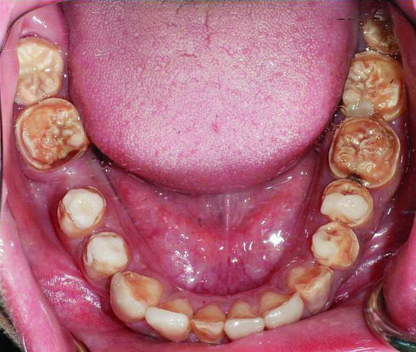
A 25-year-old female patient with amelogenesis imperfecta required restorations of her posterior teeth to improve function and eliminate tooth sensitivity. The maxillary and mandibular anterior teeth were previously restored with definitive restorations.
Intraoral and radiographic examination confirmed the diagnoses of hypocalcified type of amelogenesis imperfecta and acquired tooth loss of the mandibular right first molar (Figures 5 and 6) (Gadhia, et al., 2012).

Figure 5: Preoperative occlusal view of the maxillary teeth
Figure 6: Preoperative occlusal view of the mandibular teeth
The treatment plan involved adhesive onlays to restore the maxillary and mandibular posterior teeth. Inadequate interocclusal space was present between the molar teeth, and the patient was not willing to accept metal restorations for the definitive prostheses. Preparation for glass ceramic onlays could have detrimental effects on the tooth vitality.
Monolithic translucent zirconia onlays on the molar teeth were planned to facilitate the rehabilitation allowing minimal tooth reduction of 0.5 mm.
The preoperative treatment planning involved articulated study models and diagnostic wax-ups (Malik, et al., 2012). After completion of the diagnostic stages of the treatment plan, the definitive restorations were constructed using monolithic translucent zirconia onlays (Straumann Cares monolithic restorations) for the molar teeth and lithium disilicate onlays (IPS e.max, Ivoclar Vivadent) for the premolar teeth (Malik, et al., 2012).
The casts were scanned with CS2 scanner and the Cares Visual software (Straumann Cares System 8.0) (Kapos and Evans, 2014).
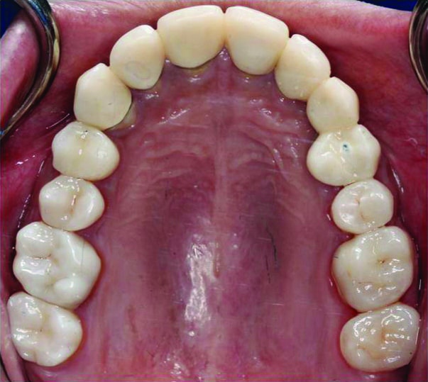
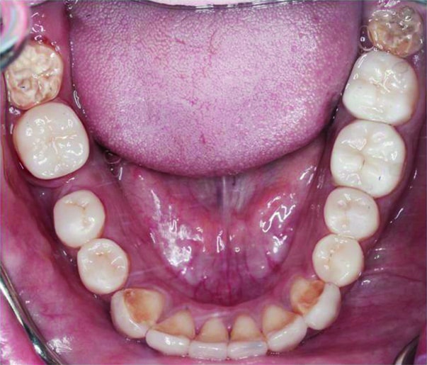
Subsequently, the restorations were stained to achieve an optimal color match (Rinke and Fischer, 2013). All restorations were cemented with a resinous cement with zirconia primer (Multilink Automix, Ivoclar Vivadent) (Figures 7 and 8) (Thompson, et al., 2011).
The patient was satisfied with the functional and esthetic outcome at the end of the treatment, tooth sensitivity was controlled, and no complications were noticed in the teeth or restorations at the 9-month review appointment.

Figure 7: Postoperative maxillary occlusal view showing the monolithic translucent zirconia restorations
Figure 8: Postoperative mandibular occlusal view showing the monolithic translucent zirconia restorations
Discussion
The literature review revealed that the long-term survival of monolithic translucent zirconia restorations lacks evidence. Nevertheless, improved properties regarding esthetics, tooth reduction, and ceramic chipping in comparison to traditional zirconia cores are reported (Nakamura, et al., 2015; Jung, et al., 2010; Harianawala, et al., 2014).
Preliminary outcomes show high survival rates for full-arch and single-unit tooth- and implant-supported prosthesis (Carames, et al., 2015; Venezia, et al., 2015; Moscovitch, 2015), but longer observation periods are necessary to draw definitive conclusions.
Traditionally, metal restorations have been used in patients with developmental conditions, especially in cases of reduced interocclusal space, which show good long-term outcomes but compromised esthetics (Gadhia, et al., 2012; Malik, et al., 2012).
Monolithic translucent zirconia restorations could offer an alternative restorative material in cases of reduced interocclusal space where esthetic requirements are critical, allowing minimal tooth reduction of 0.5 mm (Nakamura, et al., 2015).
Careful polishing after adjusting the zirconia surfaces to prevent wear to the opposing teeth and cementation with a resinous cement with zirconia primer are prerequisite to a successful outcome (Preis, et al., 2015; Rinke and Fischer, 2013).
There are limitations on the use of monolithic zirconia restorations. Their fabrication requires computer-aided design and computer-aided manufacturing (CAD/ CAM) technology and a multi-step polishing protocol after occlusal adjustment, which requires a variety of special diamond burs, diamond-impregnated silicone instruments, and diamond pastes. Besides, there is no evidence regarding the effect on the survival of the prostheses or the opposing teeth if the surface glaze wears off.
Regular review appointments and individualized maintenance are suggested to monitor the integrity of the prostheses, the condition of the abutment teeth, and to identify any complications at an early stage.
Summary
Monolithic translucent zirconia restorations may offer improved esthetics, minimal tooth reduction, and elimination of ceramic chipping compared to traditional zirconia cores and conventional metal restorations. However, there is limited evidence on the survival of monolithic translucent zirconia restorations. Preliminary outcomes suggest promising results, but longer observation periods are necessary as the use of monolithic translucent zirconia increases.
Acknowledgments
The authors would like to thank Alaa Abou Hasan (dental technician, Ceramic Studios Ltd., London) for the ceramic work and Kali Ranshi (specialty registrar in restorative dentistry, The Royal London Dental Hospital and Queen Mary University of London, Barts and The London School of Medicine and Dentistry, London, UK) for her contribution in the second clinical case toward the completion of the definitive restorations of the anterior teeth.
References
- Buser D, Halbritter S, Hart C, et al. Early implant placement with simultaneous guided bone regeneration following single-tooth extraction in the esthetic zone: 12-month results of a prospective study with 20 consecutive patients. J Periodontol. 2009;80(1):152-162.
- Carames J, Tovar Suinaga L, Yu YC, Pérez A, Kang M. Clinical advantages and limitations of monolithic zirconia restorations full arch implant supported reconstruction: case series. Int J Dent. 2015;392496
- Cardelli P, Manobianco FP, Serafini N, Murmura G, Beuer F. Full-arch, implant-supported monolithic zirconia rehabilitations: pilot clinical evaluation of wear against natural or composite teeth. J Prosthodont. 2016;25(8):629-633.
- Chen ST, Beagle J, Jensen SS, Chiapasco M, Darby I. Consensus statements and recommended clinical procedures regarding surgical techniques. Int J Oral Maxillofac Implants. 2009;24(suppl):272-278.
- Conrad HJ, Seong WJ, Pesun IJ. Current ceramic materials and systems with clinical recommendations: a systematic review. J Prosthet Dent. 2007;98(5):389-404.
- Esthetic Modifiers. In: The SAC Classification in Implant Dentistry. Chen S, Dawson A, eds. 2009; Berlin: Quintessence Publishing.
- Arkutu N, Gadhia K, McDonald S, Malik K, Currie L. Amelogenesis imperfecta: the orthodontic perspective. Br Dent J. 2012;212(10):485-489.
- Goodacre CJ, Campagni WV, Aquilino SA. Tooth preparations for complete crowns: an art form based on scientific principles. J Prosthet Dent. 2001;85(4):363-376.
- Guazzato M, Albakry M, Ringer SP, Swain MV. Strength, fracture toughness and microstructure of a selection of all-ceramic materials. Part I. Pressable and alumina glass-infiltrated ceramics. Dent Mater. 2004;20(5):441-448.
- Guazzato M, Albakry M, Ringer SP, Swain MV. Strength, fracture toughness and microstructure of a selection of all-ceramic materials. Part II. Zirconia-based dental ceramics. Dent Mater. 2004;20(5):449-456.
- Hämmerle CH, Chen ST, Wilson TG. Consensus statements and recommended clinical procedures regarding the placement of implants in extraction sockets. Int J Oral Maxillofac Implants. 2004;19:26-28.
- Harianawala HH, Kheur MG, Apte SK, Kale BB, Sethi TS, Kheur SM. Comparative analysis of transmittance for different types of commercially available zirconia and lithium disilicate materials. J Adv Prosthodont. 2014;6(6):456-661.
- Heffernan MJ, Aquilino SA, Diaz-Arnold AM, Haselton DR, Stanford CM, Vargas MA. Relative translucency of six all-ceramic systems. Part II: core and veneer materials. J Prosthet Dent. 2002;88(1): 10-15.
- Jemt T. Restoring the gingival contour by means of provisional resin crowns after single-implant treatment. Int J Periodontics Restorative Dent. 1999;19(1):20-29.
- Johansson C, Kmet G, Rivera J, Larsson C, Vult Von Steyern P. Fracture strength of monolithic all-ceramic crowns made of high translucent yttrium oxide-stabilized zirconium dioxide compared to porcelain-veneered crowns and lithium disilicate crowns. Acta Odontol Scand. 2014;72(2):145-153.
- Jung YS, Lee JW, Choi YJ, Ahn JS, Shin SW, Huh JB. A study on the in-vitro wear of the natural tooth structure by opposing zirconia or dental porcelain. J Adv Prosthodont. 2010;2(3):111-115.
- Kapos T, Evans C. CAD/CAM technology for implant abutments, crowns, and superstructures. Int J Oral Maxillofac Implants. 2014;29(suppl):117-136.
- Larsson C, Wennerberg A. The clinical success of zirconia-based crowns: a systematic review. Int J Prosthodont. 2014;27(1):33-43.
- Le M, Papia E, Larsson C. The clinical success of tooth and implant-supported zirconia-based fixed dental prostheses. A systematic review. J Oral Rehabil. 2015;42(6):467-480.
- Lewis S, Parel S, Faulkner R. Provisional implant-supported fixed restorations. Int J Oral Maxillofac Implants. 1995;10(3):319-325.
- Malik K, Gadhia K, Arkutu N, McDonald S, Blair F. The interdisciplinary management of patients with amelogenesis imperfecta – restorative dentistry. Br Dent J. 2012;212(11):537-542.
- Martin W, Lewis E, Nicol A. Local risk factors for implant therapy. Int J Oral Maxillofac Implants. 2009;24(suppl):28-38.
- Mehl C, Harder S, Wolfart M, et al. Retrievability of implant-retained crowns following cementation. Clin Oral Implants Res. 2008;19:1304-1311.
- Mericske-Stern RD, Taylor TD, Belser U. Management of the edentulous patient. Clinical Oral Implants Research. 2000;11(suppl):108-125.
- Moscovitch MS, Saba S. The use of a provisional restoration in implant dentistry: a clinical report. Int J Oral Maxillofac Implants. 1996;11(3):395-399.
- Moscovitch M. Consecutive case series of monolithic and minimally veneered zirconia restorations on teeth and implants: up to 68 months. Int J Periodontics Restorative Dent. 2015;35(3):315-323.
- Mundhe K, Jain V, Pruthi G, Shah N. Clinical study to evaluate the wear of natural enamel antagonist to zirconia and metal ceramic crowns. J Prosthet Dent. 2015;114(3):358-363.
- Nakamura K, Harada A, Inagaki R, et al. Fracture resistance of monolithic zirconia molar crowns with reduced thickness. Acta Odontol Scand. 2015;30:1-7.
- Pjetursson BE, Sailer I, Makarov NA, Zwahlen M, Thoma DS. All-ceramic or metal-ceramic tooth-supported fixed dental prostheses (FDPs)? A systematic review of the survival and complication rates. Part II: Multiple-unit FDPs. Dent Mater. 2015;31(6):624-639.
- Preis V, Schmalzbauer M, Bougeard D, Schneider-Feyrer S, Rosentritt M. Surface properties of monolithic zirconia after dental adjustment treatments and in vitro wear simulation. J Dent. 2015; 43(1):133-139.
- Rinke S, Fischer C. Range of indications for translucent zirconia modifications: clinical and technical aspects. Quintessence Int. 2013;44(8):557-66)
- Rosentritt M, Preis V, Behr M, Hahnel S, Handel G, Kolbeck C. Two-body wear of dental porcelain and substructure oxide ceramics. Clin Oral Investig. 2012;16(3):935-943.
- Sailer I, Fehér A, Filser F, Gauckler LJ, Lüthy H, Hämmerle CH. Five-year clinical results of zirconia frameworks for posterior fixed partial dentures. Int J Prosthodont. 2007;20(4):383-388
- Sailer I, Makarov NA, Thoma DS, Zwahlen M, Pjetursson BE. All-ceramic or metal-ceramic tooth-supported fixed dental prostheses (FDPs)? A systematic review of the survival and complication rates. Part I: Single crowns (SCs). Dent Mater. 2015;31(6):603-623.
- Stawarczyk B, Ozcan M, Roos M, Trottmann A, Hämmerle CH. Fracture load and failure analysis of zirconia single crowns veneered with pressed and layered ceramics after chewing simulation. Dental Materials Journal. 2011;30(4):554-562.
- Thompson JY, Stoner BR, Piascik JR, Smith R. Adhesion/cementation to zirconia and other non-silicate ceramics: where are we now? Dent Mater. 2011;27(1):71-82.
- Venezia P, Torsello F, Cavalcanti R, D’Amato S. Retrospective analysis of 26 complete-arch implant-supported monolithic zirconia prostheses with feldspathic porcelain veneering limited to the facial surface. J Prosthet Dent. 2015;114(4):506-512.
Stay Relevant With Implant Practice US
Join our email list for CE courses and webinars, articles and mores


