Educational aims and objectives
This article aims to present an overview of the main approaches of augmenting hard tissues in implant dentistry.
Expected outcomes
Implant Practice US subscribers can answer the CE questions to earn 2 hours of CE from reading this article. Take the quiz by clicking here. Correctly answering the questions will demonstrate the reader can:
- Define the process of bone regeneration and realize some steps to a successful process.
- Identify some types of grafting materials.
- Recognize the technique for block bone grafting.
- Identify when a sinus augmentation would be necessary.
- Identify the need for socket preservation.
Dr. Amit Patel offers up a primer in the techniques available to implant dentists for successful, predictable hard tissue management
Strong foundations are needed for successful dental implant placement. However, some patients present with insufficient quality or quantity of bone to support a dental implant, which can be caused by a variety of different reasons, including tooth loss, trauma, infection, and periodontal disease.
Without sufficient healthy bone, instability and loss of a dental implant can occur, along with the functional and esthetic problems that go with it. Consequently, bone and soft tissue augmentation is often required to re-create the lost anatomy and to achieve the optimal occlusal function and esthetic results that are essential for a successful outcome (Schneider, et al., 2010).
The reconstruction of tissue and bone through surgical procedures is well published and predictable. There are various techniques that can be used (Fu and Wang, 2012) such as guided bone regeneration and bone block grafting, as well as procedures such as socket preservation and sinus augmentation — all of which can be used to great effect in a wide range of indications for both functional and esthetic purposes.
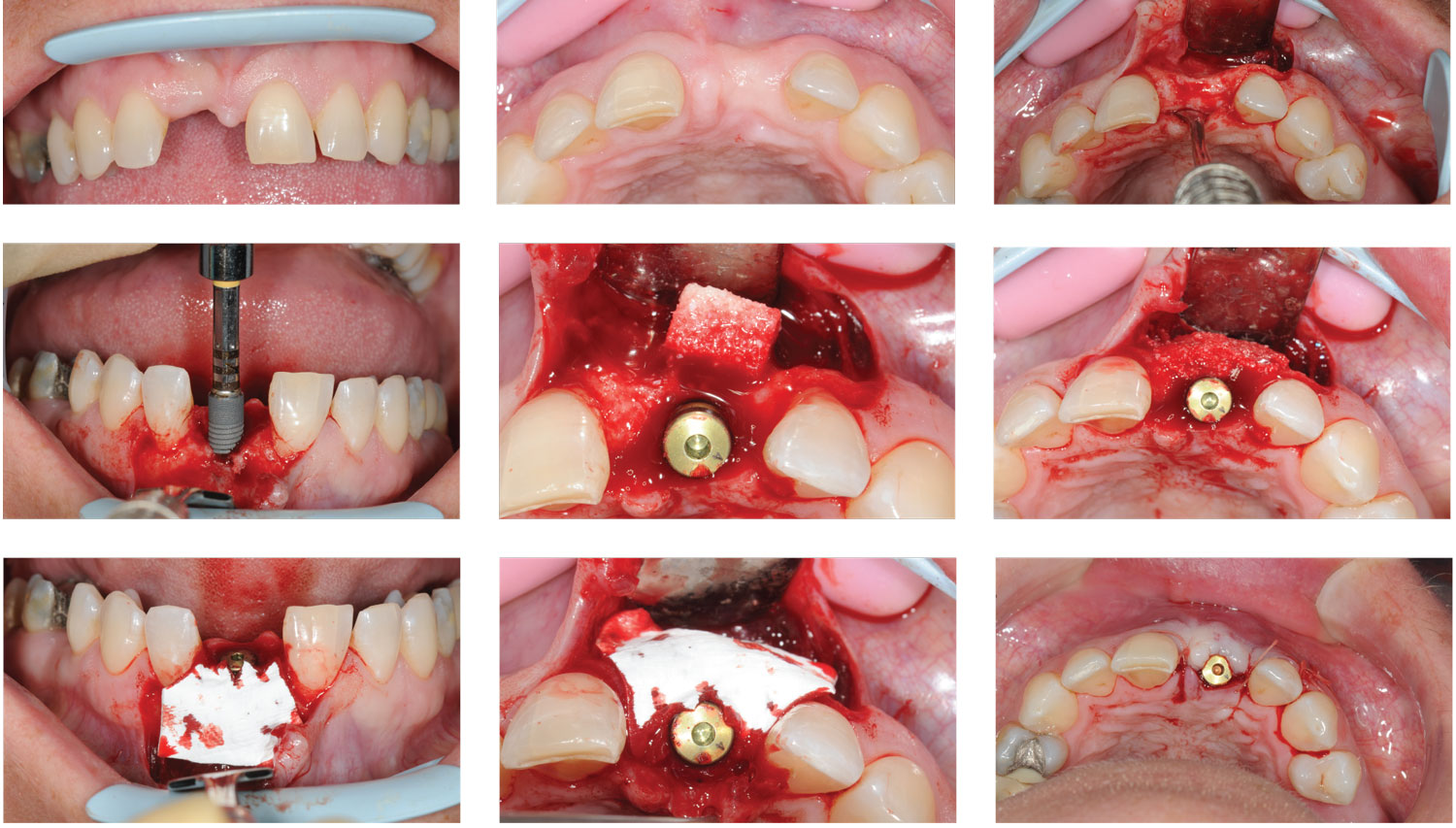
Figures 1-9: Case study 1: GBR combined with dental implant placement. One 4.2×11 mm Astra Tech Implant System™ EV implant was placed with a healing abutment. A buccal defect was present. At least 2 mm of buccal bone is required — therefore, a collagenated xenograft was placed and porcine membrane stabilized over the healing abutment utilizing the poncho technique. Figures 7-9 show the Geistlich Combi-Kit used with interrupted resorbable sutures
Guided bone regeneration (GBR)
Guided bone regeneration, or GBR, is a surgical technique where bone is regenerated using bone grafting material and a barrier membrane, maintaining a space over the grafting material into which osteogenic cells can migrate and colonize to form increased volume of bone.
It can be applied to extraction sockets, horizontal ridge augmentation, and the correction of defects around dental implants to build bone both vertically and horizontally.
Predictable bone regeneration requires a high level of technical skill, as well as a comprehensive understanding of wound healing and the following main biological principles, known collectively as “PASS.”
- P – primary wound closure to ensure undisturbed and uninterrupted wound healing
- A – angiogenesis to provide necessary blood supply and undifferentiated mesenchymal cells
- S – space maintenance/creation to facilitate adequate space for clot stability and ultimately predictable bone growth
- S – stability of wound and dental implant to induce blood clot formation and uneventful healing (Wang and Boyapati, 2006)
In some situations, GBR should be performed as an entirely separate procedure, and only when the site is sufficiently healed should dental implant surgery begin.
However, it is possible for clinicians to combine dental implant placement with bone grafting at the same time. This can reduce treatment time significantly and produce results that are difficult to achieve in other ways.
Figures 10-20: Case study 2: Sinus augmentation surgery and dental implant placement at 6 months’ post-surgery. A lateral wall approach utilizing piezosurgery was taken in this case. The thin alveolar ridge needed block grafts. Allograft blocks were trimmed to the correct shape and thickness and fixed with fixation screws. Bovine bone and porcine membrane were placed over the blocks to reduce resorption. A periosteal releasing incision was made to achieve passive closure over the graft. Horizontal mattress sutures were employed to stabilize the flap and interrupted sutures. When the site was uncovered at 6 months, a good ridge width had been reconstructed for implant placement
Grafting materials
Bone grafting materials are derived from allograft (human), xenograft (animals from another species such as bovine or porcine), or alloplast (synthetic material). They work as scaffold and stimulate the body to form natural bone at the site of the dental implant.
Barrier membranes are made from a biocompatible material and are used to promote growth and guide bone regeneration. These can be used with or without bone grafting material and protect and stabilize the bone graft. In addition, as gingival tissue grows faster than bone, the membrane effectively isolates the graft site, preventing gingival tissue from filling the area.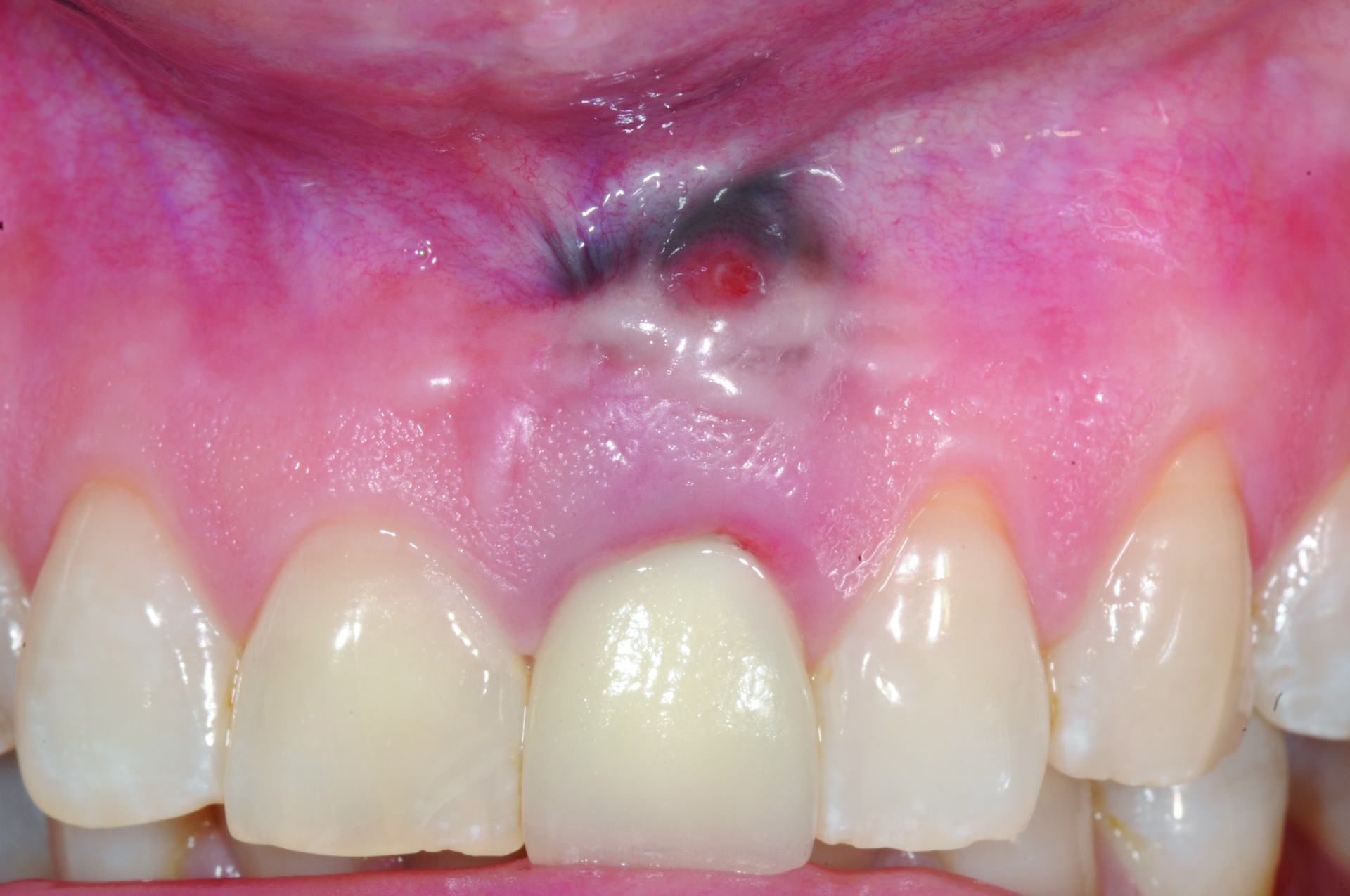


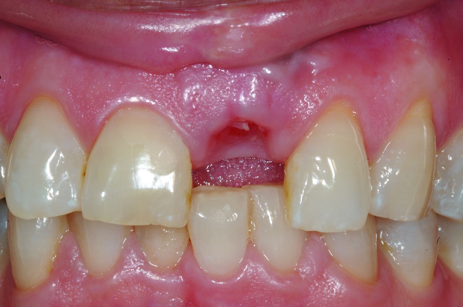
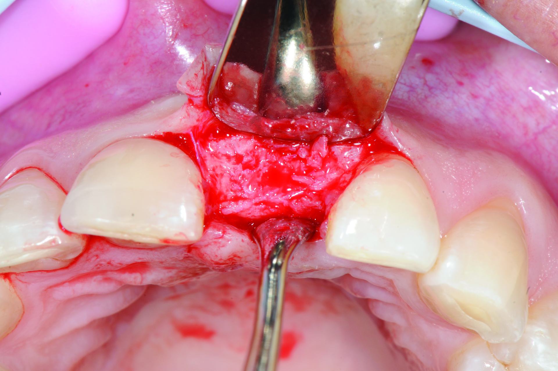
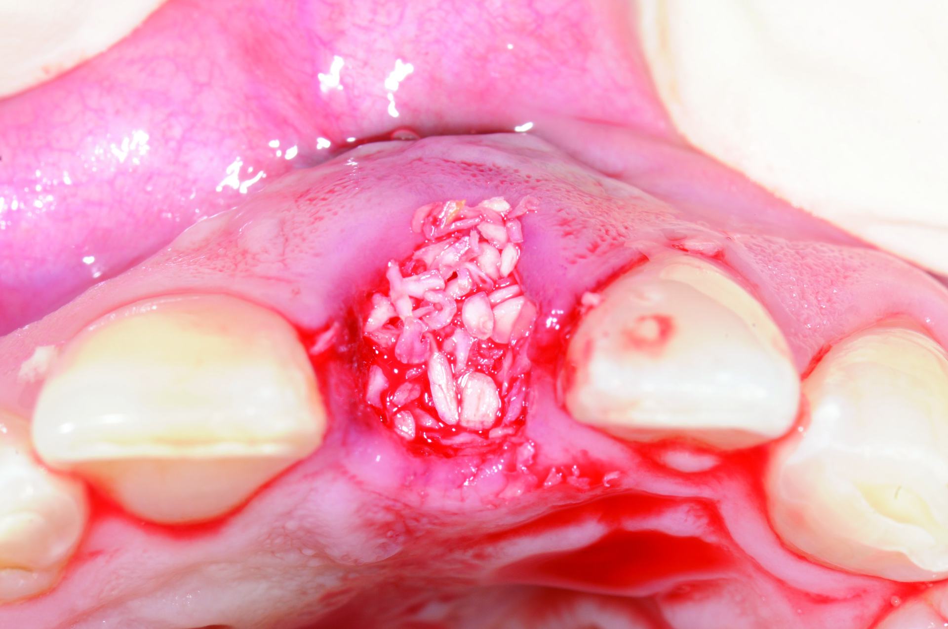

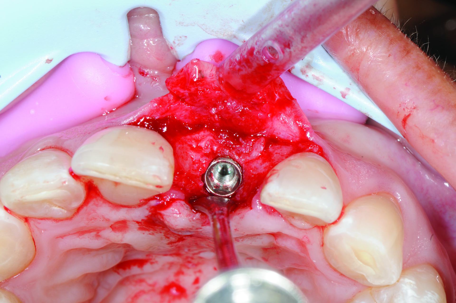 Figures 21-29: Case study 3: Socket preservation post extraction and dental implant placement at 4 months post-surgery. Periapical area draining UL1. The tooth was removed, and the socket curetted. No buccal plate was present. Allograft bone was placed into the socket and a porcine membrane placed to reconstruct the buccal plate. The site was left to heal for 4 months until uncovered for implant placement. Good bone reconstruction of the ridge was noted, and the implant placed, with no GBR needed
Block bone grafting
The technique of block bone grafting aims to restore the original anatomy using an autogenous graft (from the patient’s body), or xenogeneic or allograft normally from elsewhere in the oral cavity or from a secondary site such as the intraforamen region or the ramus in the mandible.
This grafting material is both osteoinductive and osteoconductive, which means it will induce the growth of new bone cells, and allow growth of bone cells in and around it. These unique properties generally allow for quicker bone formation, but some clinicians find this technique challenging because both the donor site and the treatment site must be managed.
However, it is very effective in cases where there is a significant defect in order to improve the integrity of the site. All materials work well, but the gold standard is to utilize human bone.
Sinus augmentation
In some patients — for example, those who have had periodontal disease or have lost molar teeth for many years — the maxillary sinuses can be positioned in close proximity to the jaw.
In these cases, it is common to complete sinus augmentation (or a sinus lift). This involves lifting the sinus membrane and placing a graft to develop bone height in order to accommodate a dental implant.
Socket preservation
Socket preservation may help reduce the bone volume and dimensional changes following tooth extraction (Ten Heggeler, et al., 2011). This procedure helps to compensate for the resorption of the facial bone wall and is particularly beneficial when dental implant placement needs to be delayed.
By preserving the socket immediately after extraction, further bone augmentation at a later date may not be necessary. In addition, reducing bone resorption and promoting bone formation in this way increases the likelihood of dental implant survival. In an experienced clinician’s hands, these techniques and many others are very effective for improving bone and tissue volume in order to achieve optimal function and esthetics with dental implants.
References
- Fu JH, Wang HL. The sandwich bone augmentation technique. Clinical Advances in Periodontics. J Periodontal. 2012;2(3):172-177.
- Schneider D, Grunder U, Ender A, Hämmerle CH, Jung RE. Volume gain and stability of peri-implant tissue following bone and soft tissue augmentation: 1-year results from a prospective cohort study. Clin Oral Implants Res. 2011;22(1):28-37.
- Ten Heggeler JM, Slot DE, Van der Weijden GA. Effect of socket preservation therapies following tooth extraction in non-molar regions in humans: a systematic review. Clin Oral Implants Res. 2011;22(8):779-788.
- Wang HL, Boyapati L. “PASS” principles for predictable bone regeneration. Implant Dent. 2006;15(1):8-17.


