Educational aims and objectives
This article aims to describe the key aspects of preparing and optimizing PRF, as well as its benefits and limitations.
Expected outcomes
Implant Practice US subscribers can answer the CE questions by taking the quiz to earn 2 hours of CE from reading this article. Correctly answering the questions will demonstrate the reader can:
Take the quiz by clicking here.
- Define the PRF preparation technique.
- Identify currently used centrifugation protocols.
- Realize techniques for optimizing the quality of PRF.
- Identify the physical handling and application of PRF.
- Recognize the benefits and limitations of PRF.
Drs. Johan Hartshorne and Howard Gluckman show the benefits and limitations of platelet-rich fibrin for tissue healing and regeneration during the implant process.
Drs. Johan Hartshorne and Howard Gluckman discuss the optimization and application of PRF — along with its benefits and limitations
Platelet-rich fibrin (PRF), a patient blood-derived living biomaterial, is increasingly being investigated and used worldwide by clinicians as an adjunctive autologous biomaterial to promote bone and soft tissue healing and regeneration. PRF technology has grabbed the attention of clinicians because it is derived from the patients’ own blood; is readily available; is easy to prepare; can be produced immediately at the chairside; is easy to use; and widely applicable in dentistry, while being financially realistic for the patient and the clinician, and with virtually no risk of a rejection reaction (foreign body response).
The 3D architecture of the fibrin matrix provides the PRF membrane with great density, elasticity, flexibility, and strength that are excellently suited for handling, manipulation, and suturing.
Optimal PRF membrane quality and treatment success is dependent on quick collection of blood and transfer to the centrifuge (2 minutes as per Choukroun); use of proper centrifugation protocol; maturation of the clot for 4 to 8 minutes before use; preparation of the membrane using a standardized preparation technique; and appropriate conservation of the membrane before use.
The PRF can be used as a membrane (A-PRF or L-PRF), liquid or injectable form (i-PRF), plug or the membrane can be cut in fragments, and applied either in stand-alone therapies (i.e., plug, filler, or protective barrier); additive therapies (i.e., added or mixed to bone substitutes); or used in combination therapies with other biomaterials (i.e., protective barrier in GBR procedures).
At present, very little is understood about PRF generated from patients with coagulation disorders or patients on medications that affect blood clotting (heparin, warfarin, or platelet inhibitors).
Its lack of rigidity and fast degradation (biodegradability) may limit its application as a sole barrier membrane in GTR procedures. One of the clinical limitations to deal with is the heterogeneity in the quality of platelets and blood components of various PRF protocols.
At this stage in time there is not a single RCT or CCT to compare the effectiveness of A-PRF or L-PRF protocols. Furthermore, in vitro studies that claim superiority or inferiority of a specific PRF preparation have yet to be validated by independent clinical trials. Standardized and optimized PRF preparation protocols and their effectiveness still have to be validated through independent robust randomized controlled clinical trials.
The purpose of this review (part 2 of 3) is to analyze the available literature on PRF relating to:
- PRF preparation technique
- Optimizing the quality of PRF
- The physical handling and application of PRF
- The benefits and limitations of PRF
An improved understanding of these aspects will facilitate the clinicians’ ability to enhance the therapeutic applications of PRF in the fields of implant dentistry, periodontology, and oral surgery.

Introduction
The prospect of having new therapies, biomaterials, and bioactive surgical additives available that will improve success and predictability of patient outcomes in soft and bone tissue healing and regeneration are key treatment objectives in dental implantology, periodontology, and oral surgery.
PRF, a patient blood-derived and autogenous living biomaterial, is increasingly being investigated and used worldwide by clinicians as an adjunctive autologous biomaterial to promote bone and soft tissue healing and regeneration. The gold standard for in vivo tissue healing and regeneration requires the mutual interaction between a scaffold (fibrin matrix), platelets, growth factors, leukocytes, and stem cells (Kawase, 2015).

These key elements are all active components of PRF, and when combined and prepared properly are involved in the key processes of tissue healing and regeneration, including cell proliferation and differentiation, extracellular matrix synthesis, chemotaxis and angiogenesis (neo-vascularization) (Dohan, et al., 2012; 2014).
An improved understanding of the development, biological, and physiological properties and characteristics of PRF in tissue healing and regeneration over the past 2 decades has led to more successful therapeutic applications, especially in the fields of implant dentistry, periodontology, and oral surgery.
Methodology, search strategy, and inclusion criteria
An electronic MEDLINE®/PubMed® and Google Scholar search was performed for all articles on platelet-rich fibrin (PRF) and platelet concentrates up to May 2016. The search was complemented by an additional hand search of selected journals in oral implantology, oral surgery, and periodontology, as well as gray literature. The reference lists and bibliographies of all included publications were also screened for relevant studies. The search was limited to the English language.
Randomized controlled trials (RCTs), controlled clinical trials (CCTs), case reports, case series, prospective, retrospective, and in vitro/in vivo studies were included in the narrative review. Animal studies were excluded from this review.
How is PRF prepared, and how can I use it?
Blood drawing
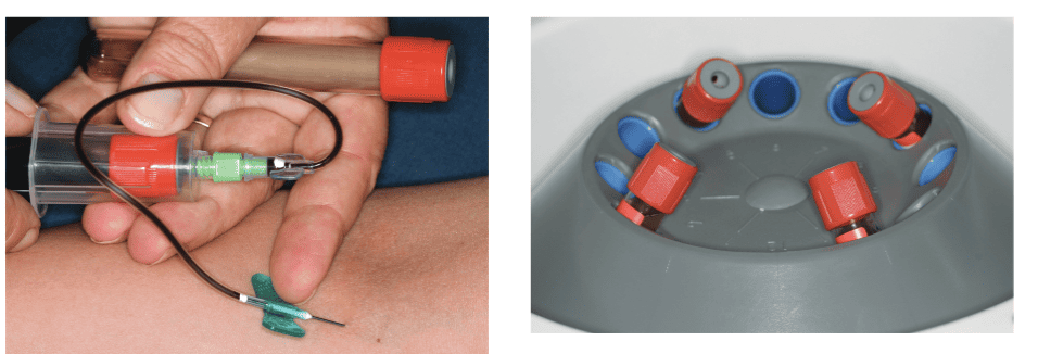
A major advantage of PRF is that it has a simple preparation protocol. Blood is drawn from the patient using a sterile 10 ml vacutainer (two to 12 tubes) just before or during surgery (Figure 1).
Dental clinicians have the following options for drawing blood:
- Doing it themselves (venepuncture courses are available)
- Asking an anesthetist or sedationist when using general anesthesia or conscious sedation for procedures
- Contracting the services of a qualified nurse
The tubes with collected blood samples are immediately (within 2 minutes after collecting the blood sample) placed in the centrifuge and processed using a single centrifugation step. The clinical success of the PRF protocol is dependent on a quick collection of blood and its transfer to the centrifuge because blood will automatically start to coagulate after 1-2 minutes and make it difficult to obtain the required clot quality (Ali, et al., 2016). Failure to accomplish the quick preparation of PRF could cause a diffuse polymerization of fibrin, which is not ideal for tissue healing.
Centrifugation
The tubes should always be balanced by opposing two tubes to equilibrate the centrifugation forces and to prevent vibrations during the centrifugation process (Figure 2).
At the end of the centrifugation spin, the caps for A-PRF or L-PRF (not i-PRF) are removed and the tubes placed in a sterile tube holder (Figure 3). The blood sample with the clot is allowed to rest/mature for approximately 4-8 minutes before extracting the clot from the tube. (Figure 4)
The centrifugation process activates the coagulation process and separates the blood sample into three different layers: an acellular plasma at the top of the tube; a strongly polymerized fibrin clot is formed in the middle; and blood cells (red corpuscle base) are gathered at the bottom of the tube.4,5,6
Currently used centrifugation protocols
There are various centrifugation processing protocols that are currently being used.
- Original Choukroun’s PRF protocol (standard protocol): 3,000 rpm/10 minutes
- Dohan Ehrenfest’s group (leukocyte- and platelet-rich fibrin [L-PRF]): 2,700 rpm/12 minutes
- Choukroun’s advanced PRF (A-PRF),enriched with leukocytes: 1,300 rpm/ 8 minutes
- Choukroun’s i-PRF (solution/gel): 700 rpm/3 minutes
Effect of centrifugation protocols on the optimum fibrin clot:cell ratio
Current data shows that there is a differential distribution of red blood cells, platelets, and leukocytes in the PRF clot depending on the centrifugal force used (Hauser, et al., 2013). The clinical efficacy of different centrifugation protocols, however, still needs to be independently validated with controlled clinical trials.
In vitro studies showed that a longer centrifugation protocol (2,700 rpm) produces a denser (stronger) fibrin clot with less inter-fibrous space containing less cells compared to the shorter centrifugation protocol of A-PRF (1300 rpm) that produced a less dense fibrin clot with a looser inter-fibrous structure containing more cells (Hauser, et al., 2013).
Dohan and co-workers (2010) found in their in vitro studies that the original L-PRF protocol produces larger clots and membranes, and a more intense release of growth factors than the modified A-PRF protocol. Based on the findings of their studies they suggested that centrifuge characteristics and protocols may have a very significant impact on the cell, growth factors, and fibrin architecture of a PRF clot and membrane.
In contrast, another recent in vivo study showed that Choukroun’s new formulation of PRF (A-PRF) had a more gradual release of growth factors, up to a 10-day period and stimulated significantly higher growth factor release over time when compared to Choukroun’s standard PRF (Öncu and Alaaddinoõglu 2015).
The latter investigators concluded that A-PRF may prove clinically beneficial for future regenerative procedures. The clinical effectiveness and implications of above-mentioned findings still have to be validated through robust randomized controlled clinical trials.
Effect of test tube type and compression on clot quality
Research data suggests that the type of vacutube that is used (i.e., dry glass or glass-coated plastic tubes) and the compression process of the clot (forcible or soft) do not seem to influence the architecture of this autologous biomaterial.
However, both parameters could influence the growth-factor content and the matrix properties of the product (Tatullo, et al., 2012). The influence of these preparation factors requires further analysis, and their effect on the clinical efficacy needs to be validated with good quality clinical trials.
Preparation of PRF membranes
Each fibrin clot concentrates most platelets (97%) and more than half of the leukocytes from a 9 ml blood harvest (Tatullo, et al., 2012, Clipet, et al., 2012, Kanayama, et al., 2016).
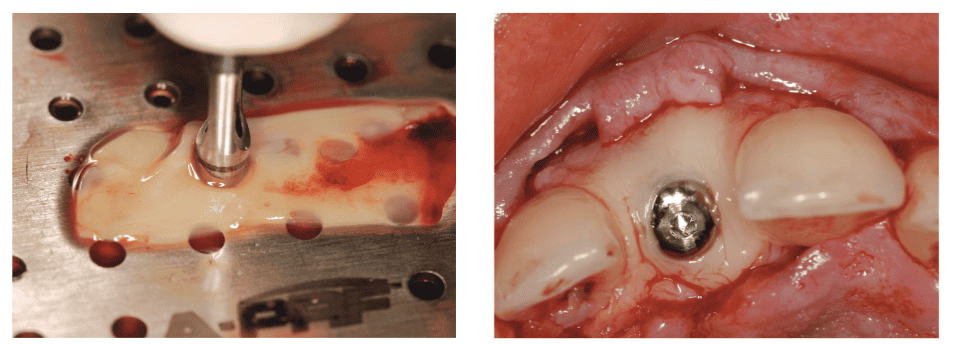
The PRF clot is removed from the tube with a sterile tweezer. The fibrin clot is separated from the red blood cell fragment, approximately 2 mm below the dividing line, using a scissor (Figure 5).
The section of the blood clot attached to the fibrin clot contains the stem cells. The PRF clots are placed in the Box grid (Process, France) or Xpression™ Kit (Intra-Lock, Boca Raton, Florida) and covered with the lid. The PRF membranes are ready for use after 2 minutes.
It provides a three-dimensional matrix of platelets, leukocytes, and growth factors.
A PRF membrane remains usable many hours after preparation, as long as the PRF is prepared correctly and conserved in physiologic conditions.
Moreover, the use of the PRF Box, a user-friendly and inexpensive tool, allows for standardized preparation of homogeneous PRF membranes with a higher growth factor content, avoids the dehydration or death of the leukocytes living in the PRF clot, and also prevents the shrinkage of the fibrin matrix architecture (Mazor, et al., 2009; Simonpieri, et al., 2011; Tajim, et al., 2013).
The concept of “the optimal PRF scaffold or composites,” tailored for specific clinical applications, is still in a process of virtual development. Further clinical trials are required to validate this concept, and what centrifugal force and time is required to get the optimum PRF composite biomaterial for specific clinical applications.
At this stage in time there is not a single RCT or CCT to compare the effectiveness of any of the above-mentioned PRF (A-PRF or L-PRF) protocols. Furthermore, in vitro studies that claim superiority or inferiority of a specific PRF preparation have not been validated by independent clinical trials. Therefore, based on these facts, no preference or distinction is made between any of the above-mentioned PRF preparation protocols (A-PRF or L-PRF) in this review.
Practical guidelines for optimizing the quality of PRF
PRF is a living biomaterial that requires a good knowledge and skills on how to produce, prepare, conserve, and use it most effectively and efficiently (Ali, et al., 2016; Tatullo, et al., 2012; Zhang, et al., 2012). Incorrect use could lead to damaged, dry product leading to inconsistent clinical results (Bölükbas, et al., 2013). The most critical factor affecting the success of PRF in healing and regenerative therapy is the quality of PRF preparations.
Following are some practical guidelines how to optimize the quality of PRF, increase clinical efficiency and consistency, and how to prevent common mistakes.
Limit centrifuge vibration during PRF preparation
Centrifuge rotational speed and subsequent vibrations have been shown to have a direct impact on the architecture and cell content of a PRF clot (Diss, et al., 2008).
The Dohan group have hypothesized that the type/make of centrifuge machine has an effect on rotational speed and vibrations. The latter, however, has not been validated with clinical trials.
Vibrations can be limited through following the following rules:
- Always make sure that the centrifuge tubes are filled equally (1 cm from the top).
- Always balance the rotor properly. Every tube must have a balance or opposing tube (Figure 2).
- Do not balance with a vacutube filled with water: The distribution of the densities will be incorrect and cause unnecessary vibration.
- If properly balanced and used, the rotor should accelerate smoothly and with a constant change in the pitch of the motor sound.
- Any vibrations or unusual sounds should cause the cessation of operation immediately by the operator.
- Initial vibrations with start of centrifugation can be reduced by holding your hand on the lid.
- Never leave the centrifuge until you are certain that it has reached its operating speed and is functioning properly. All rotors go through a minor vibration phase when they first start. There will be a minor flutter when the rotor reaches this vibration point — do not confuse this with a serious vibration caused by imbalance.
Patients receiving anticoagulants
At present, very little is understood about PRF generated from patients with coagulation disorders or patients on medications that affect blood clotting (heparin, warfarin, or platelet inhibitors). When patients are on any type of anticoagulant therapy, they have a longer coagulation time, therefore, it is suggested to centrifuge blood for longer periods, or increase the waiting time after centrifugation (approximately 5-10 minutes). However, there is no data available to support this recommendation.
Standardized and efficient preparation of PRF
The PRF box (Box grid or Xpression Kit) was designed to collect and transform up to 16 PRF clots into membranes in sterile conditions and to conserve them in a clean and wet environment before use (Figure 6A). The box also contains compression wells and maces to compress the PRF clots into dense PRF cylinders, easy to use for filling cavities (such as extraction sockets).
It is suggested that gentle pressure is used to prevent squeezing out all of the plasma contained in the original PRF clots (Toffler, et al., 2010). Compressing the clot too hard or too long results in shrinkage of the fibrin network, release of growth factors, dehydration, and damage of leukocyte content (Simonpieri, et al., 2011). PRF membranes or plugs are ready for use within 2 minutes after compression of clots in the Box grid (Figure 6B) or plug well (cylinder).
The serum exudate collected in the bottom of the box can be used for a longer conservation of the membranes and is ready to be mixed with a bone biomaterial for grafting. This standardized approach also allows an increase in the total growth factors release of the PRF membrane itself (Simonpieri, et al., 2011).
The exudate in the bottom of the box is rich in proteins (Vitronectin and Fibronectin). This solution can be recovered with a syringe and used to hydrate biomaterials, flush the surgical site, wet the implant surface, and to preserve harvested autogenous bone blocks, rather than using saline.
Conservation of the PRF membrane
PRF is a blood-derived living tissue and must be handled carefully to keep its cellular content alive and stable. Conserve the PRF clot in its centrifugation tube. As long as the serum has not been flushed away from the clot, the growth factor content remains stable. It is a good way to gain 5 to 15 minutes (Simonpieri, et al., 2011). PRF membranes remain usable many hours after preparation, as long as the PRF is prepared correctly and conserved in physiologic conditions (Clipet, et al., 2012; Simonpieri, et al., 2011; Tajim, et al., 2013).
The PRF Box, a user-friendly and inexpensive tool, guarantees the adequate preparation of homogeneous PRF membranes with a higher growth factor content, avoids dehydration or death of the leukocytes living in the PRF clot, and also prevents shrinkage of the fibrin matrix architecture.
Optimal preparation and selection
For the standardization of PRF preparations and to preserve platelets and growth factors, it is suggested not to squeeze out all of the plasma contained in the original PRF clots (Toffler, et al., 2010). Platelets are not equally distributed inside and on the surface of the PRF clot. Therefore, in a clinical situation, when growth factors provided by platelets are expected and desired, the platelet-rich region adjacent to the red thrombus should be used (Toffler, et al., 2010).
Therefore, always place the part of the clot closest to the thrombus closest to the grafting site. This part of the clot contains the highest concentration of platelets and stem cells required for bone regeneration. Thus, it is necessary to preserve a small RBC layer at the PRF clot end to collect as many platelets and leukocytes as possible. This part of the procedure is done with scissors and remains operator-dependent (Bölükbas, et al., 2013).
Optimizing and preserving growth factor release
For the standardization of PRF preparations as a grafting material for tissue regeneration, PRF membranes should always be preserved in a wet serum environment (Toffler, et al., 2010). The compression procedure of the clots into membranes is performed with a gentle, slow, and homogeneous pressure to prevent squeezing out all the serum contained in the original PRF clot (Bölükbas, et al., 2013; Toffler, et al., 2010; Kanayama, et al., 2014).
This will ensure that the final membrane always remains homogeneously wet and soaked with serum. This gentle method thus avoids extracting and losing a significant amount of extrinsic-incorporated platelet growth factors (Clipet, et al., 2012). Significant amounts of growth factors are released during the first 20 minutes after preparation (Mazor, et al., 2009). PRF membranes should therefore be used as quickly as possible after preparation.
However, this release is considerably less significant when forcible extraction is avoided. In physiologic conditions, the main release occurs after several hours (Mazor, et al., 2009), and PRF can be used a long time after preparation, as long as the material is conserved in the adequate conditions.
Handling the PRF: clinical application
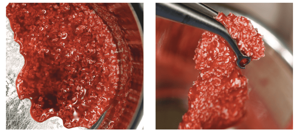
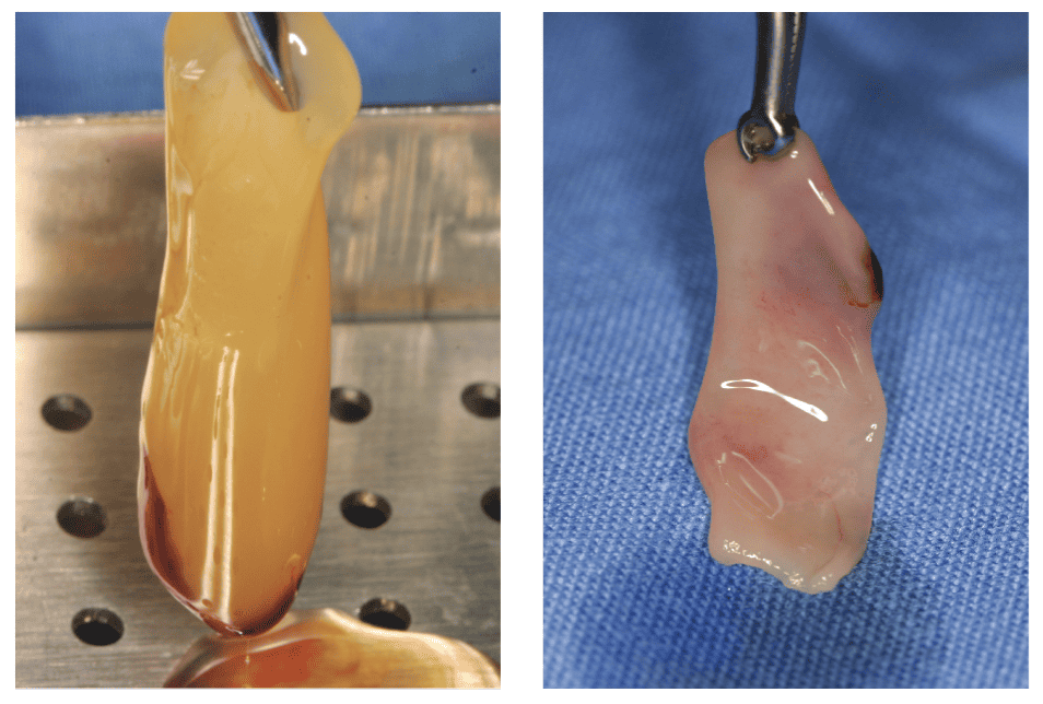
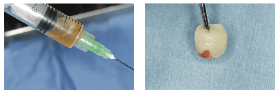
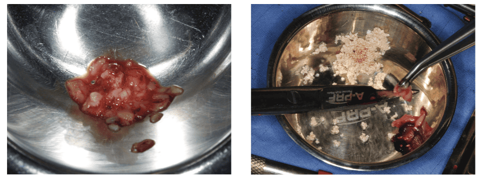
PRF membranes are easy to drape over a surgical or augmented site. The elastic consistency of the PRF membrane also allows the clinician to punch a hole in the membrane to drape over a healing abutment before suturing the flap (Ghetiu, et al., 2015) (Figures 7A and 7B). Mixing autogenous bone or bone substitutes (allografts) with i-PRF (PRF lquid) for use in GBR procedures transforms particulate bone into an easy to handle gel consistency (Figures 8A and 8B).
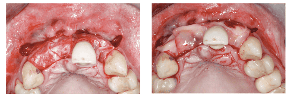
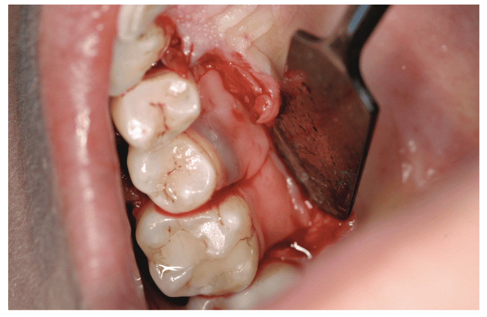
PRF liquid (i-PRF) can be injected above, or PRF (A-PRF or L-PRF) membrane placed above the GBR or GTR membrane (Figures 9A and 9B) to act as an interposition barrier to protect and stimulate the bone compartment, and as a healing membrane in order to improve the soft tissue healing and remodeling, and thus avoid soft tissue dehiscence (Peck, et al., 2011; Inchingolo, et al., 2010).
Using PRF: physical application
PRF (A-PRF or L-PRF) can either be used as a clot (Figure 10A), membrane (Figure 10B), injectable liquid (i-PRF) (Figure 10C), plug (Figure 10D), or the membrane can be cut up in fragments (Figures 10E and 10F). PRF can either be applied in stand-alone, additive, or in combination therapies.
Stand-alone therapies
Typical stand-alone therapies include using the fibrin plug or membrane as a filler material in extraction sockets (Figure 11) to prevent complications and to enhance socket healing (Choukroun, et al., 2006).
PRF can also be used as a protective barrier membrane to seal off and promote healing of oroantral communications following extractions (Magremanne, et al., 2009); to close a palatal connective tissue harvesting site (Zhao, et al., 2011); or as sole grafting material in sinus floor elevations (Kanayama, et al., 2014; Del Corso, et al., 2012; Rao, et al., 2013; Öncü and Erbeyo 2015; Simonpieri, et al., 2009; Simonpieri, et al., 2009) or as a vestibuloplasty wound bandage (Figure 12).

Additive therapies
PRF (membrane or liquid) can be added or mixed to bone substitutes (Figure 13) such as xenograft or biphasic calcium phosphate (BCP) to enhance the formation of new bone (Toeroek and Dohan 2013). Research suggests that bone healing is more effective when PRF is mixed with autogenous bone or bone substitutes such as DBBG and BCP in bone augmentation or GBR procedures (Vijayalakshmi 2012). However, there is limited evidence to support this as a clinical guideline.
Combination therapies
The PRF membrane is typically used in combination with other biomaterials in bone augmentation and grafting sites as a graft material or barrier membrane (Hamzacebi, et al., 2015) (Figures 9A and 9B). The purpose of PRF is to activate and facilitate the healing and regenerative capacity of the host tissue, by providing a strong fibrin scaffold, major growth factors, and allowing space for tissue regeneration. Using PRF as a protective barrier on bone graft sites helps avoid perforations of the weakened gingival tissues and prevent associated contamination of the bone graft below.
Key biological function of a PRF membrane
PRF membranes are not comparable to heterologous resorbable collagen or non-resorbable (i.e., dPTFE) membranes. PRF membranes belong to a completely different category of membrane, namely natural autologous membrane (Shilbli, et al., 2013). A PRF membrane is as natural as the host tissue, while heterologous membranes are considered as foreign bodies by the host tissues and interfere with the natural tissue healing process. A PRF membrane can be used for three purposes:
Bioactive barrier
A PRF membrane is a blood clot prepared in an optimized form that is rich in cells and growth factors, and acts as a natural bioactive barrier, allowing interaction with the tissues below and above it. This interaction with tissues facilitates natural tissue regeneration (NTR) and healing (Inchingolo, et al., 2010). PRF will undergo quicker remodeling (biodegradation) in situ than a resorbable collagen membrane, but will also promote a strong induction on the periosteum/gingival tissue due to the slow release of growth factors and other matrix proteins (Krasny, et al., 2011; Kim, et al., 2013).
Competitive interposition barrier
GTR membranes are cell-proof barriers against soft tissue invagination, whereas PRF membranes allow cells to migrate through it, thus allowing new blood vessel formation that will facilitate regenerative and healing interactions between the tissues below and above the PRF membrane. The PRF membrane is a highly stimulating matrix, attracting cell migration and differentiation preferentially, and also reinforcing the natural periosteal barrier.
The hard and soft tissues migrate and interact within the PRF matrix. The PRF matrix becomes the interface between the tissues and therefore avoids the migration of the soft tissues deeper within grafted defect or augmented site. This biological characteristic is referred to as a competitive barrier (Krasny, et al., 2011). However, it is important to recognize that using PRF as a competitive barrier does not have the graft stability or space maintenance characteristics of a normal collagen membrane, and therefore cannot be recommended for use as such.
Protective barrier and healing booster
PRF membranes are frequently used for the protection of the grafted area and as a healing booster for the soft tissues above the grafted defects or augmented sites (Inchingolo, et al., 2010). The purpose of the PRF membrane is not only to protect the blood clot and/or the graft material, like in the GTR concept, but also to promote the induction of a strong and thick periosteum and gingiva. This boosted periosteum functions as a true barrier between the soft tissue and bone compartments, and constitutes probably the best protection and regenerative barrier for the intrabony defects (Inchingolo, et al., 2010) (Figure 14).
What are the benefits of PRF?
PRF has increasingly grabbed the attention of clinicians worldwide because this biomaterial is of natural origin (autologous/derived from the patients’ own blood); can be produced chairside; is easy to make, readily available; easy to use within the daily clinical routine; widely applicable in dentistry, while being financially realistic for the patient and the health care system (Joseph, et al., 2014).
The following major advantages support the clinical use of PRF in tissue repair and regeneration processes.
Natural (autologous) biomaterial
Regenerative therapies are now shifting away from using allogenic and xenogeneic biomaterials to autologous biomaterials. While other membranes are considered as foreign bodies by the host tissues and interfere with the natural tissue healing process, a PRF membrane is as natural as the host tissue with virtually no risk of infection, immune or a rejection reaction (foreign body response) (Hauser, et al., 2013; Kanayama, et al., 2014; Barone, et al., 2014).
Easy and efficient to use
Preparing PRF is easy, fast, and user-friendly to use within the daily clinical routine (Kanayama, et al., 2014; Inchingolo, et al., 2010; Nacopoulos, et al., 2014; Del Corso and Dohan 2013; Shah, et al., 2014). PRF is prepared on-site (in the clinical setting) simply by drawing blood from the patient for immediate use, thus reducing patient waiting time. Currently used protocols for the preparation of autologous PRF are standardized and easy for clinicians and clinical assistants to use. Furthermore, there is no limitation on quantity of PRF membranes required.
In comparison with a natural blood clot, a PRF membrane is a solid material, has a strong fibrin architecture, and easier to handle and to position (Kanayama, et al., 2014; Barone, et al., 2014). Fibrin three-dimensional architecture provides the PRF membrane with great density, elasticity, flexibility, and strength (Ali, et al., 2016; Del Corso and Dohan 2013) that is better suited for manipulation and suturing (Hauser, et al., 2013). The elastic consistency of the PRF membrane allows the clinician to punch it to allow for placing it around a healing or prosthetic abutment or even suture it.
Increased clinical performance
Recent studies have shown that, compared with previous generation PRP, PRF exhibits a greater expression and concentration of growth factors and matrix proteins, which are released more slowly due to the three-dimensional architecture of the adhesive glycoproteins in the fibrin, which results in significantly better performance (Mazor, et al., 2009; Simonpieri, et al., 2011; Gamal, et al., 2016).
Moreover, A-PRF shows a more gradual release of growth factors, up to a 10-day period, and stimulated significantly higher growth factor release over time when compared to Choukroun’s standard PRF (Öncu and Alaaddinoõglu 2015).
Safety
Blood is drawn from the patient, and there is therefore reduced donor site morbidity. PRF rarely causes complications such as membrane exposure; an unwanted outcome that has been observed in cases using biodegradable barrier membranes (Shah, et al., 2015). A further advantage of PRF is the extremely low risk of infection. Moreover, no in vitro cytotoxicity effects have been detected whatever the quantity of PRF used (Sharma and Pradeep 2011; 2011; Pradeep, et al., 2012).
Increased healing potential
PRF increases the predictability of wound healing and regeneration potential of tissues (Zhao, et al., 2011; Bansal and Bharti 2013; Thorat 2011; Pradeep, et al., 2012; Troiano et al 2016; Pradeep and Sharma 2011; Gupta, et al., 2014; Panda, et al., 2014).
Reduced morbidity
A clinical advantage of PRF as a graft material is related to avoidance of a donor site and risk of morbidity, thus resulting in a decrease in patient discomfort, post-surgical pain, and bleeding after operation (Kanayama, et al., 2014; Panda, et al., 2014). PRF is not only a platelet concentrate but also an “immune node” that is able to stimulate defense mechanisms.
It is even likely that the significant inflammatory regulation noted on surgical sites treated with PRF is the outcome of retro-control effects from cytokines trapped in the fibrin network and released during the remodeling of this initial matrix (Kanayama, et al., 2016). Furthermore, evidence suggests that the content of platelet alpha granules might have a bactericidal effect, mediated by molecules called thrombocidines that may have an important contribution toward reducing postoperative infections (Lekovic, et al., 2012; Joseph, et al., 2012). The anti-hemorrhagic properties of PRF are also advantageous and convenient during surgical procedures (Ghetiu, et al., 2015).
Cost-benefit ratio
PRF has an outstanding potential cost-to-benefit ratio. A platelet-rich fibrin (PRF) membrane is a readily available and inexpensive biomaterial that is beneficial in implant dentistry, oral surgery, and periodontal procedures (Bajaj, et al., 2013).
The ease of preparation and cost-effectiveness of PRF membrane offers a huge advantage over other commercially available membranes (Patil, et al., 2014). It is widely applicable in dentistry, while being financially realistic for the patient and the healthcare system. PRF is currently the safest and most economical choice for patients and clinicians for improving healing and tissue (soft tissue and bone) regeneration outcomes.
It can spare the patient from additional operating field (second-surgery morbidity) and also save cost through avoiding use of alloplastic or xenogenic membranes and reduce the quantity of synthetic grafting materials (Thorat 2011). It is also an economical alternative to expensive recombinant growth factors when used in conjunction with osseous grafts.
Affordability and cost-effectiveness
PRF is a simple and inexpensive biomaterial to use (Kanayama, et al., 2014; Shah, et al., 2014). The initial investment for producing PRF preparations is potentially affordable for most private practices to invest in (Simonpieri, et al., 2011).
No contraindications
PRF membranes have no contraindications: they can be used in all kinds of patients, especially in patients with systemic conditions where healing is compromised (i.e., diabetics and smokers), or in surgically compromised situations (damaged flap). In these situations, PRF will promote soft tissue healing and stimulate the healing of a damaged flap and reduce the risks of flap necrosis after a surgery. It is a common point that all fibrin-based products (platelet concentrates) are frequently used for the stimulation of angiogenesis and to reduce the risk of flap necrosis in many general surgery applications (Ranganathan and Chandran 2014; Desarda, et al., 2013).
Open access and widely applicable
The PRF technique is open access and thus can be widely developed and used in private practice without commercial considerations (Inchingolo, et al., 2010). The clinical applications of PRF in other fields of dentistry, such as endodontics, are also increasing exponentially.
Limitations
The rapid use of PRF without delay or short handling time may be a potential limitation. Lack of rigidity and fast degradation (biodegradability) (Sambhav, et al., 2014; Simonpieri, et al., 2011; Dohan 2009) may limit its application in GTR procedures.
PRF can be considered a healing bio-material that can be utilized in regenerative surgical procedures to fasten healing, but its application as a barrier membrane in GBR is doubtful due to its poor mechanical properties (Patil, et al., 2014).
Owing to the fact that PRF is an autologous product, the availability of this bio-material in larger amounts is also a concern. One of the clinical limitations to deal with is the heterogeneity in the quality of platelets and blood components. At present, very little is understood about PRF generated from patients with coagulation disorders or patients on medications that affect blood clotting (heparin, warfarin, or platelet inhibitors).
Conclusion
The therapeutic use of PRF for accelerating tissue healing and regeneration has increasingly grabbed the attention of clinicians worldwide because this biomaterial is of natural origin (autologous/derived from the patients’ own blood); PRF technology is readily available; is easy to prepare; can be produced immediately at chairside; is easy to use; and widely applicable in dentistry, while being financially realistic for the patient and the clinician, and with virtually no risk of a rejection reaction (foreign body response). The three-dimensional architecture of fibrin provides the PRF membrane with great density, elasticity, flexibility, and strength that are excellently suited for handling, manipulation, and suturing.
Optimal PRF membrane quality and treatment success is dependent on quick collection of blood and transfer to the centrifuge; use of proper centrifugation protocol; maturation of the clot for 5 minutes before use; preparation of the membrane using a standardized preparation technique; and appropriate conservation of the membrane before use.
The PRF can be used as a membrane (A-PRF or L-PRF), gel (i-PRF), plug, or the membrane can be cut in fragments and applied either in stand-alone therapies (i.e., plug, filler, or protective barrier); additive therapies (i.e., added or mixed to bone substitutes); or used in combination therapies with other biomaterials (i.e., protective barrier in GBR procedures).
More importantly, the use of PRF enables local delivery of a fibrin matrix, cells, growth factors, and proteins that provide unique biological properties and cues for promoting new blood vessel formation, and potentially accelerating wound healing and tissue regeneration, while at the same time reducing adverse events. Consequently, the benefits of PRF in wound and bone healing, its antibacterial and antihemorrhagic effects, the low risks with its use, and the availability of easy and low-cost preparation methods should encourage more clinicians to adopt this technology in their practices for the benefit of their patients
One of the clinical limitations to deal with is the heterogeneity in the quality and quantity of platelets and blood components due to use of different PRF preparation protocols. At this point in time there is not a single RCT or CCT to compare the effectiveness of any of the PRF (A-PRF or L-PRF) protocols. Furthermore, in vitro studies that claim superiority of inferiority of a specific PRF preparation have not been validated by independent clinical trials.
PRF preparation protocols (A-PRF and L-PRF) and the clinical effectiveness thereof still have to be validated and standardized through independent robust randomized controlled clinical trials.
Remember this is part 2 of this very informative series on platelet-rich fibrin! Read part 1 again or for the first time (and subscribers remember to take the 2-credit quiz) here.
References
- Agarwal K, Chandra C, Agarwal K, Kumar N. Lateral sliding bridge flap technique along with platelet-rich fibrin and guided tissue regeneration for root coverage. J Indian Soc Periodontol. 2013;17:801-805.
- Aleksić Z, Janković S, Dimitrijević B, et al. The use of platelet-rich fibrin membrane in gingival recession treatment. Srp Arh Celok Lek. 2010;38:118.
- Ali S, Bakry SA, Abd-Elhakam H. Platelet-rich fibrin in maxillary sinus augmentation: a systematic review. J Oral Implantol. 2015;41(6):746-753.
- Aroca S, Keglevich T, Barbieri B, Gera I, Etienne D. Clinical evaluation of a modified coronally advanced flap alone or in combination with a platelet-rich fibrin membrane for the treatment of adjacent multiple gingival recessions: a 6-month study. J Periodontol. 2009;80(2):244-252.
- Bajaj P, Pradeep AR, Agarwal E, et al. Comparative evaluation of autologous platelet-rich fibrin and platelet-rich plasma in the treatment of mandibular degree II furcation defects: a randomized controlled clinical trial. J Periodont Res. 2013;48(5):573-581.
- Barone A, Ricci M, Romanos GE, Tonelli P, Alfonsi F, Covani U. Buccal bone deficiency in fresh extraction sockets: a prospective single cohort study. Clin Oral Implants Res. 2015;26(7):823-830.
- Bansal C, Bharti V. Evaluation of efficacy of autologous platelet-rich fibrin with demineralized freeze dried bone allograft in the treatment of periodontal intrabony defects. J Indian Soc Periodontol. 2013;7(3):361-366.
- Bolukbasi N, Ersanli S, Keklikoglu N, Basegmez C, Ozdemir T. Sinus augmentation with platelet-rich fibrin in combination with bovine bone graft versus bovine bone graft in combination with collagen membrane. J Oral Implantol. 2015;41(5):586-595.
- Boora P, Rathee M, Bhoria M. Effect of Platelet Rich Fibrin (PRF) on Peri-implant Soft Tissue and Crestal Bone in One-Stage ImplantPlacement: A Randomized Controlled Trial. J Clin Diagn Res. 2015;9(4):ZC18-ZC21.
- Choukroun J, Diss A, Simonpieri A, et al. Platelet-rich fibrin (PRF): a second-generation platelet concentrate. Part V: histologic evaluations of PRF effects on bone allograft maturation in sinus lift. Oral Surg Oral Med Oral Pathol Oral Radiol Endod. 101:299-303.
- Choukroun J, Diss A, Simonpieri A, et al. Platelet-rich fibrin (PRF): a second-generation platelet concentrate. Part IV: clinical effects on tissue healing. Oral Surg Oral Med Oral Pathol Oral Radiol Endod. 2006;101(3):e56-e60
- Clipet F, Tricot S, Alno N, et al. In vitro effects of Choukroun’s platelet-rich fibrin conditioned medium on 3 different cell lines implicated in dental implantology. Implant Dent. 2012;21:51-56.
- Del Corso M, Sammartino G, Dohan EDM. Clinical evaluation of a modified coronally advanced flap alone or in combination with a platelet-rich fibrin membrane for the treatment of adjacent multiple gingival recessions: a 6-month study. J Periodontol. 2009;80(11):1694-1697.
- Del Corso M, Vervelle A, Simonpieri A, et al. Current knowledge and perspectives for the use of platelet-rich plasma (PRP) and platelet-rich fibrin (PRF) in oral and maxillofacial surgery part 1: Periodontal and dentoalveolar surgery. Curr Pharm Biotechnol. 2012;13(7):1207-1230.
- Del Corso M, Dohan EDM. Immediate implantation and peri-implant Natural Bone Regeneration (NBR) in the severely resorbed posterior mandible using Leukocyte- and Platelet-Rich Fibrin (L-PRF): a 4-year follow-up. POSEIDO. 2013;1(2):109-116.
- Desarda HM, Gurav AN, Gaikwad SP, Inamdar SP. Platelet rich fibrin: a new hope for regeneration in aggressive periodontitis patients: report of two cases. Indian J Dent Res. 2013;24:627-630.
- Diss A, Dohan DM, Mouhyi J, Mahler P. Osteotome sinus floor elevation using Choukroun’s platelet-rich fibrin as grafting material: a 1-year prospective pilot study with micro threaded implants. Oral Surg Oral Med Oral Pathol Oral Radiol Endod. 2008;105:572-579.
- Dohan EDM, de Peppo GM, Doglioli P, Sammartino G. Slow release of growth factors and thrombospondin-1 in Choukroun’s platelet-rich fibrin (PRF): a gold standard to achieve for all surgical platelet concentrates technologies. Growth Factors. 2009;27(1):63-69.
- Dohan EDM. How to optimize the preparation of leukocyte- and platelet-rich fibrin (L-PRF, Choukroun’s technique) clots and membranes: introducing the PRF Box. Oral Surg Oral Med Oral Pathol Oral Radiol Endod. 2010;110(3):275-278; author reply 8-80
- Dohan EDM, Bielecki T, Mishra A, Borzini P, Inchingolo F, Sammartino G, Rasmusson L, Evert PA. In search of a consensus terminology in the field of platelet concentrates for surgical use: platelet-rich plasma (PRP), platelet-rich fibrin (PRF), fibrin gel polymerization and leukocytes. Curr Pharm Biotechnol. 2012;13(7):1131-1137.
- Dohan EDM, Andia I, Zumstein M, Zhang C-Q, Nelson R. Pinto NR, Bielecki T. Classification of platelet concentrates (Platelet-Rich Plasma-PRP, Platelet- Rich Fibrin-PRF) for topical and infiltrative use in orthopedic and sports medicine: current consensus, clinical implications and perspectives. Muscles Ligaments Tendons J. 2014;4(1):3-9.
- Eren G, Atilla G. Platelet-rich fibrin in the treatment of localized gingival recessions: a split-mouth randomized clinical trial. Clin Oral Invest. 2014;18(8):1941-1948
- Gamal AY, Abdel Ghaffar KA, Alghezwy OA. Crevicular Fluid Growth Factors Release Profile Following the Use of Platelets Rich Fibrin (PRF) and Plasma Rich Growth Factors (PRGF) in Treating Periodontal Intrabony Defects (Randomized Clinical Trial). J Periodontol. 2016;87(6):654-662.
- Ghetiu A, Sirbu D, Topalo V, et al. Tissue engineering with Platelet-Rich-Fibrin in Oral region. Clin Oral Impl Res. 2015;26(suppl 12):205
- Gupta SJ, Jhingran R, Gupta V, et al. Efficacy of platelet-rich fibrin vs. enamel matrix derivative in the treatment of periodontal intrabony defects: a clinical and cone beam computed tomography study. J Int Acad Periodontol. 2014;16(3):86-96.
- Gupta G, Puri K, Bansal M, Khatri M, Kumar A. Platelet-Rich Fibrin-Reinforced Vestibular Incision Sub periosteal Tunnel Access Technique for Recession Coverage. Clin Adv Periodontics. 2015;5:248-253.
- Hamzacebi B, Oduncuoglo B, Alaaddinogl EE. Treatment of peri-implant bone defects with platelet-rich fibrin. Int J Periodont Restorative Dent. 2015;35(3):415-422.
- Hauser F, Gaydarov N, Badoud I, et al. Clinical and histological evaluation of postextraction platelet-rich fibrin socket filling: a prospective randomized controlled study. Implant Dent. 2013;22(3):295-303.
- Inchingolo F, Tatullo M, Marrelli M, et al. Trial with Platelet-Rich Fibrin and Bio-Oss used as grafting materials in the treatment of the severe maxillary bone atrophy: clinical and radiological evaluations. Eur Rev Med Pharmacol Sci. 2010;14(12):1075-1084.
- Jankovic S, Aleksic Z, Milinkovic I, Dimitrijevic B. The coronally advanced flap in combination with platelet-rich fibrin (PRF) and enamel matrix derivative in the treatment of gingival recession: a comparative study. Eur J Esthet Dent. 2010;5:260-273.
- Jankovic S, Aleksic Z, Klokkevold P, et al. Use of platelet-rich fibrin membrane following treatment of gingival recession: a randomized clinical trial. Int J Periodontics Restor Dent. 2012;32(5):41-50.
- Joseph RV, Raghunath A, Sharma N. Clinical effectiveness of autologous platelet rich fibrin in the management of infrabony periodontal defects. Singapore Dent J. 2012;33(1):5-12.
- Joseph VR, Sam G, Amol NV. Clinical evaluation of autologous platelet rich fibrin in horizontal alveolar bony defects. J Clin Diagn Res. 2014;8(11) ZC43-ZC47.
- Kanayama T, Sigetomi T, Sato H, Yokoi M. Crestal approach sinus floor elevation in atrophic posterior maxilla using only platelet rich fibrin as grafting material: A computed tomography evaluation of 2 cases. J Oral Maxillofac Surg Med Path. 2014;26(4):519-525.
- Kanayama T, Horii K, Senga Y, Shibuya Y. Crestal Approach to Sinus Floor Elevation for Atrophic Maxilla Using Platelet-Rich Fibrin as the Only Grafting Material: A 1-Year Prospective Study. Implant Dent. 2016;25(1):32-38.
- Kawase T. Platelet-rich plasma and its derivatives as promising bioactive materials for regenerative medicine: basic principles and concepts underlying recent advances. Odontology. 2015;103(2):126-135.
- Keceli HG, Kamak G, Erdemir EO, Evginer MS, Dolgun A. The Adjunctive Effect of Platelet-Rich Fibrin to Connective Tissue Graft in the Treatment of Buccal Recession Defects: Results of a Randomized, Parallel-Group Contolled Trial. J Periodontol. 2015;86(11):1221-1230.
- Kim JS, Jeong MH, Jo JH, Kim SG, Oh JS. Clinical application of platelet-rich fibrin by the application of the Double J technique during implant placement in alveolar bone defect areas: case reports. Implant Dent. 2013;22(3):244-249.
- Krasny K, Kaminski A, Krasny M, et al. Clinical use of allogeneic bone granulates to reconstruct maxillary and mandibular alveolar processes. Transplant Proc. 2011;43(8):3142-3144.
- Lekovic V, Milinkovic I, Aleksic Z, et al. Platelet-rich fibrin and bovine porous bone mineral vs. platelet-rich fibrin in the treatment of intrabony periodontal defects. J Periodontal Res. 2011;47:409-417.
- Magremanne M, Baeyens W, Awada S, Vervaet C. Solitary bone cyst of the mandible and platelet rich fibrin (PRF). Rev Stomatol Chir Maxillofac. 2009;110(2):105-108.
- Mazor Z, Horowitz RA, Del Corso M, et al. Sinus floor augmentation with simultaneous implant placement using Choukroun’s platelet-rich fibrin as the sole grafting material: a radiologic and histologic study at 6 months. J Periodontol. 2009;80:2056-2064.
- Moraschini V, dos Santos EBP. Use of Platelet-Rich Fibrin Membrane in the Treatment of Gingival Recession: a Systematic Review and Meta-Analysis. J Periodontol. 2016;87(3):281-290.
- Nacopoulos C, Dontas I, Lelovas P, et al.. Enhancement of bone regeneration with the combination of platelet-rich fibrin and synthetic graft. J Craniofac Surg. 2014;25(6):2164-2168.
- Öncü EA, Erbeyoğlu AA. Enhancement of immediate implant stability and recovery by platelet-rich fibrin. Clin Oral Impl Res. 2017;27. doi: 10.11607/prd.2505
- Panda S, Jayakumar ND, Sankari M, Varghese SS, Kumar DS. Platelet rich fibrin and xenograft in treatment of intrabony defect. Contemp Clin Dent. 2014;5(4):550-554.
- Panda S, Ramamoorthi S, Jayakumar ND, Sankari M, Varghese SS. Platelet rich fibrin and alloplast in the treatment of intrabony defect. J Pharm Bioallied Sci. 2014;6:127-131.
- Patil PA, Kumar R V, Kripal K. Role and Efficacy of L- PRF matrix in the Regeneration of Periodontal Defect: A New Perspective. J Clin Diagn Res. 2014;8:ZD03-ZD05
- Peck MT, Marnewick J, Stephen L. Alveolar ridge preservation using leukocyte and platelet-rich fibrin: a report of a case. Case Rep Dent. 2011; 345048
- Platelet-Rich Fibrin Combined With a Porous Hydroxyapatite Graft for the Treatment of 3-Wall Intrabony Defects in Chronic Periodontitis: A Randomized Controlled Clinical Trial. J Periodontol. 2011;82(12):1288-1296.
- Pradeep AR, Rao NS, Agarwal E, Bajaj P, Kumari M, Naik SB. Comparative evaluation of autologous platelet-rich fibrin and platelet-rich plasma in the treatment of 3-wall intrabony defects in chronic periodontitis: a randomized controlled clinical trial. J Periodontol. 2012;83(12):1499-1507.
- Pradeep AR, Bajaj P, Rao NS, Agarwal E, Naik SB. Platelet-Rich Fibrin Combined With a Porous Hydroxyapatite Graft for the Treatment of 3-Wall Intrabony Defects in Chronic Periodontitis: A Randomized Controlled Clinical Trial. J Periodontol. 2017;88(12):1288-1296.
- Rajaram V, Thyegarajan R, Balachandran A, Aari G, Kanakamedala A. Platelet Rich Fibrin in double lateral sliding bridge flap procedure for gingival recession coverage: An original study. J Indian Soc Periodontol. 2015;19(6):665-670.
- Ranganathan AT, Chandran CR. Platelet-rich fibrin in the treatment of periodontal bone defects. J Contemp Dent Pract. 2014;15(3):372-375.
- Rao SG, Bhat P, Nagesh KS, et al. Bone Regeneration in Extraction Sockets with Autologous Platelet Rich Fibrin Gel. J Maxillofac Oral Surg. 2013;12(1):11-16.
- Sambhav J, Rohit R, Ranjana M, Shalabh M. Platelet rich fibrin (PRF) and -tricalcium phosphate with coronally advanced flap for the management of grade-II furcation defect. Ethiop J Health Sci. 2014;24(3):269-272.
- Shah M, Deshpande N, Bharwani A, et al. Effectiveness of autologous platelet-rich fibrin in the treatment of intra-bony defects: A systematic review and meta-analysis. J Indian Soc Periodontol. 2014;18(6):698-704.
- Shah M, Patel J, Dave D, Shah S. Comparative evaluation of platelet-rich fibrin with demineralized freeze-dried bone allograft in periodontal infrabony defects: A randomized controlled clinical study. J Indian Soc Periodontol. 2015;19(1):56-60.
- Sharma A, Pradeep AR. Treatment of 3-wall intrabony defects in patients with chronic periodontitis with autologous platelet-rich fibrin: A randomized controlled clinical trial. J Periodontol. 2011;82(12):1705-1712.
- Sharma A, Pradeep AR. Autologous platelet-rich fibrin in the treatment of mandibular degree II furcation defects: a randomised clinical trial. J Periodontol. 2011;82:1396-1403.
- Shilbli JA, Blay A, Tunchel S, Roth L, Nardagam G, Cassoni A, Rodriques JA. Treatment of peri-implantitis using L-PRF and ER,CR:YSGG laser in the esthetic zone: Longitudinal aspects. In: Complex situations on Implant Dentistry: Specialized Clinical Solutions. Eds: EHO Rosetti and WC Bonachela. VM Vultural, Sao Paulo; 2013.
- Simonpieri A, Del Corso M, Sammartino G, Dohan Ehrenfest DM. The relevance of Choukroun’s platelet-rich fibrin and metronidazole during complex maxillary rehabilitations using bone allograft. Part I: a new grafting protocol. Implant Dent. 2009;18(2):102-111.
- Simonpieri A, Del Corso M, Sammartino G, Dohan Ehrenfest DM. The relevance of Choukroun’s platelet-rich fibrin and metronidazole during complex maxillary rehabilitations using bone allograft. Part II: implant surgery, prosthodontics, and survival. Implant Dent. 2009;18(3):220-229.
- Simonpieri A, Choukroun J, Del Corso M, Sammartino G, Dohan EDM. Simultaneous sinus-lift and implantation using micro threaded implants and leukocyte- and platelet-rich fibrin as sole grafting material: a six-year experience. Implant Dent. 2011;20(1):2-12.
- Tajima N, Ohba S, Sawase T, Asahina I. Evaluation of sinus floor augmentation with simultaneous implant placement using platelet- rich fibrin as sole grafting material. Int J Oral Maxillofac Implants. 2013;28:77-83.
- Tatullo M, Marrelli M, Cassetta M, Pacifici A, Stefanelli LV, Scacco S, Dipalma G, Pacifici L, Inchingolo F. Platelet Rich Fibrin (P.R.F.) in reconstructive surgery of atrophied maxillary bones: clinical and histological evaluations. Int J Med Sci. 2012;9:872-880.
- Thorat MK, Pradeep AR, Pallavi B. Clinical effect of autologous platelet-rich fibrin in the treatment of intra-bony defects: a controlled clinical trial. J Clin Periodontol. 2011;38:925-932.
- Toeroek R, Dohan EDM. The concept of Screw- Guided Bone Regeneration (S-GBR). Part 3: Fast Screw- Guided Bone Regeneration (FS-GBR) in the severely resorbed preimplant posterior mandible using allograft and Leukocyte- and Platelet-Rich Fibrin (L PRF): a 4-year follow-up. POSEIDO. 2013;1(2):93-100.
- Toffler M, Toscano N, Holtzclaw D. Osteotome-mediated sinus floor elevation using only platelet-rich fibrin: an early report on 110 patients. Implant Dent. 2010;19:447-456.
- Troiano G, Laino L, Dioguardi M, et al. Mandibular Class II Furcation Defects Treatment: Effects of the Addition of Platelet Concentrates to Open Flap. A Systematic Review and Meta-analysis of RCT. J Periodontol. 2016; 87(9):1030-1038.
- Vijayalakshmi R, Rajmohan CS, Deepalakshmi D, Sivakami G. Use of platelet rich fibrin in a fenestration defect around an implant. J Indian Soc Periodontol. 2012;16(1):108-112.
- Zhang Y, Tangl S, Huber CD, Lin Y, Qiu L, Rausch-Fan X. Effects of Choukroun’s platelet-rich fibrin on bone regeneration in combination with deproteinized bovine bone mineral in maxillary sinus augmentation: a histological and histomorphometric study. J Craniomaxillofac Surg. 2012;40(4):321-328.
- Zhao JH, Tsai CH, Chang YC. Clinical and histologic evaluations of healing in an extraction socket filled with platelet-rich fibrin. J Dent Sci. 2011;6(2):116-122.


