In the first part of a series of articles, Drs. Sajid Jivraj, Mamaly Reshad, and Winston Chee look at the diagnostic factors that affect the predictability of peri-implant esthetics
Achieving esthetics with implant restorations is significantly more challenging than with conventional restorations. Diagnosis and appropriate treatment planning are critical in obtaining a successful outcome. Many manufacturers will identify their systems as esthetic — from an objective perspective, components in and of themselves are not esthetic. There is not a single component available that would be the ideal replacement for a maxillary central incisor. Esthetic outcomes are based on many variables. It is not the specific implant design, surface characteristics, or type of abutment that will guarantee an esthetic result. It is the time spent on data collection in reaching a correct diagnosis that pays dividends in terms of function and esthetics (Sullivan, 2001).

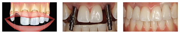
Root form cylindrical implants placed following surgical techniques described by Branemark, et al., have proven to be a predictable method for anchoring replacement teeth to the jaw bone (Branemark, et al., 1990; Naert, et al., 1992). Today, clinicians can prescribe the use of implants with the knowledge and confidence that they will predictably integrate into the jawbone. The successful integration of an implant, however, is not sufficient to declare success; implants placed in poor restorative positions result in unesthetic restorations that provide little satisfaction for the clinician or the patient. Figures 1-3 demonstrate the complexity of implant use in esthetic zones and the importance of proper treatment planning prior to implant placement.
Providing an esthetic outcome requires understanding of the objective and subjective criteria related to hard and soft tissue esthetics (Belser, 1982). Both dental and gingival esthetics act together to provide a smile with harmony and balance. The clinician must be aware of parameters related to gingival morphology, form and dimension, characterization, surface texture, and color (Magne, Belser, 2002) [Figure 4].
Ceramists can often produce restorations to match adjacent teeth in terms of color. However, if the surrounding tissues are not reconstructed, an esthetic outcome is not likely (Figures 5A and 5B). The ultimate aim is for the implant restoration to harmonize into the frame of the smile, face and, more importantly, the individual.
Treatment planning must address hard and soft tissue deficiencies and combine this with precision in implant placement; only with this approach can implant restorations be indistinguishable from the adjacent teeth (Figure 6).

Recreating what nature provided can be a formidable challenge. The physiology of wound healing after tooth extraction creates an unfavorable soft tissue complex. The remaining mucosa often recedes palatally and apically. Often this results in a restoration that appears long, and this is compounded by the absence of interdental papilla (Figure 7).
The predictability of the esthetic outcome of an implant restoration is dependent on many variables including, but not limited to:
- Patient selection and smile line
- Tooth position
- Root position of the adjacent teeth
- Biotype of the periodontium and tooth shape
- The bony anatomy of the implant site
- The position of the implant.
Patient selection and smile line
Patients who are candidates for replacement of an anterior tooth with an implant-supported restoration must understand its benefits. They must also understand the additional length of time required for treatment and additional costs that will be incurred. The clinician must also understand the patient’s desires. In most cases, the patient’s primary demand is an esthetic tooth replacement. With this in mind, it is important to establish sound clinical concepts with clearly defined parameters that lead to successful esthetics with long-term stability of the peri-implant tissues.
The major indication for a single tooth implant restoration is preservation (nonpreparation) of one or more of the adjacent teeth (Figure 8), and reduction in the rate of alveolar resorption. Additional indications would be restoration of a missing tooth to maintain a diastema and preservation of extensive fixed restorations that are intact.
A patient’s esthetic expectations must also be evaluated together with his/her lip activity and lip length. In an average smile, 75-100% of the maxillary incisors and the interproximal gingiva are displayed. In a high smile line, additional gingival tissue is exposed. Less than 75% of the incisors are exposed in a low smile line (Figures 9-11) [Tjan, Miller, The, 1984]. The clinician should be aware that the patient who presents with unacceptable tooth health, shade, or position may not give a full smile when asked. Previous photographs may aid in determining the natural position of the patient’s lip when smiling.
A high smile line poses considerable challenges when planning for implant-supported restorations in the esthetic zone because the restoration and gingival tissues are completely displayed. In these types of clinical situations, maximal efforts towards maintaining peri-implant tissue support throughout the planning, provisional, surgical, and restorative phases will be required.
The low smile line is a less critical situation because the implant restoration interface will be hidden behind the upper lip. However, this cannot be assumed, and the patient’s input must be sought to confirm this.

Tooth position
The tooth needs to be evaluated in three planes of space: apicocoronal, faciolingual, and mesiodistal. The existing tooth position will significantly influence the presenting gingival architecture. In many instances, teeth with a poor prognosis are thoughtlessly extracted. These teeth can significantly influence both the hard and soft tissue configuration.
Apico-coronal
On assessment of the apico-coronal position of the tooth, it may be more apical, more coronal, or ideal, and mimic the level of the adjacent gingival margin (Figure 12). Numerous authors have shown that following extraction and insertion of an ovate pontic, there is likely to be up to 2 mm of gingival recession, and on extraction and placement of an implant immediately the migration of the gingival margin is likely to approximate 1 mm (Kois, 1998; Saadoun, et al., 1999).

The implication of this is that if there is a hopeless tooth positioned ideally or apically, and this is extracted, the gingival margin is likely to migrate apically. Restoratively, long clinical crowns, pink porcelain, or visible metal margins will compromise the esthetic outcome. These teeth can benefit from orthodontic extrusion (Figure 13) prior to extraction, which will serve to position the gingival level at a more harmonious level (Kois, 2004; Salama, Salama, Kelly, 1996).
Faciolingual
In this dimension, the tooth position may present with different concerns. The tooth may be positioned too far facially; this often results in very thin or nonexistent labial bone. These teeth are not good candidates for orthodontic extrusion because of inadequate underlying bone. Extraction of these teeth results in significant vertical bone loss and collapse of the gingival architecture. This type of situation would benefit from bone augmentation procedures prior to implant placement. A tooth positioned more lingually would benefit from the presence of an increased amount of facial bone. This situation is more favorable prior to extraction since the resultant discrepancy in the facial free gingival margin may be minimal (Kois, 2004).
Mesiodistal
The proximity of the adjacent teeth necessary to provide proximal support and volume of interdental papillae should be evaluated. Ideally, the mesiodistal tooth width should be equal to that of the contra lateral tooth so that an esthetic outcome can be achieved (Figures 14 and 15). Excess or deficiencies in this dimension should be addressed through the use of orthodontics, enameloplasty, or restorations. For patients with diastemas, it is imperative that the decision to maintain or close the space be made prior to implant placement. If the patient refuses the above options to close the space and insists on closing the space with the implant restoration, there is a likelihood that a black triangle may ensue. This results from inadequate support from the adjacent tooth to maintain the papilla. It is important that the clinician discusses this with the patient ahead of time so disappointment with the final outcome is avoided (Figures 16 and 17).

Root position of the adjacent teeth
Part of the diagnostic work for patients who need implants is a periapical radiograph, as often root position will preclude placing of implants. Many of these patients can benefit from orthodontics to reposition malposed teeth. If the patient illustrated in Figures 18 and 19 desired implant restorations to replace congenitally missing maxillary lateral incisors, orthodontic therapy would be necessary to move the roots of the cuspid and central incisor to allow room for ideal implant placement.
Teeth with root proximity also possess very little interproximal bone, and this thin bone creates a greater risk of lateral resorption, which will decrease the vertical bone height after extraction or implant placement. When teeth are present, the use of orthodontics serves as a valuable adjunct to create space. This can be advantageous for support of proximal gingival architecture (Tarnow, Cho, Wallace, 2000; Tarnow, Magner, Fletcher, 1992).
Biotype of periodontium and tooth shape
The position of the gingival tissue around a tooth is determined by the connective tissue attachment and by the bone level. Two different periodontal biotypes have been described in relation to the morphology of the interdental papilla and the osseous architecture — the thin scalloped periodontium and the thick flat periodontium (Becker, et al., 1997).

The thin scalloped periodontium, found in less than 15% of cases, is characterized by a delicate soft tissue curtain, a scalloped underlying osseous form, and often has dehiscences and fenestrations, and a reduced quantity and quality of keratinized mucosa. Generally, interproximal tissue does not completely fill the space between adjacent teeth. This form of gingiva reacts to insults by receding facially and interproximally. As recession occurs and the interroot bone resorbs, the subsequent soft tissue loss compromises the overall esthetic result (Figure 20).
The tooth form in this type exhibits a contact point towards the incisal third essentially triangular anatomic crowns and contact areas of teeth that are small faciolingually and apico-coronally. Due to extreme taper of the roots, the bone interproximally tends to be thicker.
Characteristics of the soft tissue biotype will play a prominent role in final planning for the shoulder position of the implant. A thin biotype with highly scalloped tissue will require the implant body and shoulder to be placed more palatal to mask any titanium show-through. When implants are placed toward the palate, a slightly deeper placement is required to allow for proper emergence profile.
Combining previous factors in a patient with a high lip line and a thin biotype is extremely difficult to treat. Patients who fit into these treatment categories should be made aware of the challenges involved in obtaining an esthetic result before treatment begins.
The thick flat periodontal biotype is characterized by a denser more fibrotic soft tissue curtain, a flat thicker underlying osseous form, and an increased quantity and quality of attached keratinized gingiva. This tissue often reacts to insults by pocket formation. Flat gingiva is associated with a tooth form that is more bulbous; contact areas are located more toward the middle third of the tooth primarily square anatomic crowns and contact areas that are wide faciolingually and apico-coronally (Figure 21).
The tooth morphology appears to be correlated with the soft tissue quality. The triangular tooth shape is associated with the scalloped and thin periodontium. The contact area is located in the coronal third of the crown, underlining a long and thin papilla. The square anatomic crown shape combines with a thick and flat periodontium. The contact area is located at the middle third, supporting a short and wide papilla.
Loss of interproximal tissue in the presence of a triangular tooth form will display a wider black triangle than in a situation when a square tooth is present (Figure 21). In some cases when the adjacent teeth are to be restored, the crown form can be modified prosthetically to compensate for partial interproximal tooth loss. The contact area of the prosthetic tooth is positioned more cervically, reducing the volume of the interdental space.
The presenting tooth shape will also influence the implant restoration shape. The implant restoration should mimic its contra lateral natural tooth coronal to the free gingival margin (Figure 22). However, apical to the free gingival margin, the implant restoration will not be an anatomic replica. A delicate balance must be developed that provides adequate support of the gingival architecture, yet does not provide excessive pressure. Ideally, the facial contour should be slightly flatter than the contra lateral natural tooth to minimize apical displacement of the free gingival margin after insertion (Figure 23) [Phillips, Kois, 1998].
Parts of this article were reprinted with permission from the British Dental Journal.
Belser UC. Esthetic checklist for the fixed prosthesis. Part II: Biscuit bake try-in. In: Schärer P,Rinn LA, Kopp FR, eds. Esthetic guidelines for restorative dentistry. Chicago, IL: Quintessence; 1982:188-192.
Becker W, Ochsenbein C, Tibbetts L, Becker BE. Alveolar bone anatomic profiles as measured from dry skulls. Clinical ramifications. J Clin Periodontol. 1997;24(10):727-731.
Adell R, Eriksson B, Lekholm U, Brånemark PI, Jemt T. Long term follow-up study of osseointegrated implants in the treatment of totally edentulous jaws. Int J Oral Maxillofac Implants. 1990;5(4):347-359.
Kois JC. Esthetic extraction site development: The biological variables. Contemp Esthet Restorative Pract. 1998;2:10-18.
Kois JC. Predictable single tooth peri-implant esthetics: five diagnostic keys. Compend Contin Educ Dent. 2004;25(11):895-896, 898, 900, 906-907.
Magne P, Belser U. Natural oral esthetics. In: Bonded porcelain restorations in the anterior dentition: a biomimetic approach. Chicago, IL: Quintessence; 2002:57-99.
Naert I, Quirynen M, van Steenberghe D, Darius P. A study of 589 consecutive implants supporting complete fixed prostheses. Part II: Prosthetic aspects. J Prosthet Dent. 1992;68(6):949-956.
Phillips K, Kois JC. Aesthetic peri-implant site development. The restorative connection. Dent Clin North Am. 1998;42(1):57-70.
Saadoun AP, LeGall M, Touati B. Selection and ideal tridimensional implant position for soft tissue esthetics. Pract Periodontics Aesthet Dent. 1999;11(9):1063-1072, 1074.
Salama H, Salama M, Kelly J. The orthodontic-periodontal connection in implant site development. Pract Periodontics Aesthet Dent. 1996;8(9):923-932, 934.
Sullivan RM. Perspectives on esthetics in implant dentistry. Compend Contin Educ Dent. 2001;22(8):685-692.
Tarnow DP, Cho SC, Wallace SS. The effect of inter-implant distance on the height of inter-implant bone crest. J Periodontol. 2000;71(4):546-549.
Tarnow DP, Magner AW, Fletcher P. The effect of the distance from the contact point to the crest of bone on the presence or absence of the interproximal dental papilla. J Periodontol. 1992;63(12):995-996.
Tjan AH, Miller GD, The JG. Some esthetic factors in a smile. J Prosthet Dent. 1984;51(1):24-28.
Stay Relevant With Implant Practice US
Join our email list for CE courses and webinars, articles and mores

